Author(s):
Suraj Wasudeo Nagre1* and K N Bhosle22
1Associate Professor, Department of C.V.T.S, Grant Medical College, Mumbai, India
2Professor and Head of Department C.V.T.S, Grant Medical College, Mumbai, India
Received: 09 October, 2017; Accepted: 18 October, 2017; Published: 20 October, 2017
Suraj Wasudeo Nagre, Associate Professor, Department of C.V.T.S, Grant Medical College, Mumbai, India, E-mail:
Nagre SW, Bhosle KN (2017) Ministernotomy Thymectomy in Mysthania Gravis-Future. J Cardiovasc Med Cardiol 4(4): 070-074. 10.17352/2455-2976.000053
© 2017 Nagre SW, et al. This is an open-access article distributed under the terms of the Creative Commons Attribution License, which permits unrestricted use, distribution, and reproduction in any medium, provided the original author and source are credited.
A thymectomy is the surgical removal of the thymus gland. The thymus has been demonstrated to play a role in the development of MG. It is removed in an effort to improve the weakness caused by MG, and to remove a thymoma if present.About 10% of MG patients have a tumor of the thymus called a thymoma. Most of these tumors are benign and tend to grow very slowly; on occasion they are malignant (“cancerous”).A thymectomy is recommended for patients under the age of 60 (occasionally older) with moderate to severe MG weakness. It is sometimes recommended for patients with relatively mild weakness, especially if there is weakness of the respiratory (breathing) or oropharyngeal (swallowing) muscles. It is also recommended for all patients with a thymoma. A thymectomy is usually not recommended for patients with weakness limited to the eye muscles (ocular myasthenia gravis). The neurological goals of a thymectomy are significant improvement in the patient’s weakness, reduction in the medications being employed, and ideally eventually a permanent remission (complete elimination of all weakness off all medications). There are three basic surgical approaches transternal, transcervical and videoscopic[VATS] thymectomy each with several variations. Regardless of the technique employed, the surgical goal is to remove the entire thymus. Many believe this should include removal of the adjacent fat; others are less sure.Here we give our study report of comparision between full sternotomy against ministernotomy thymectomy patients preopt, intraopt and postopt factors, fifteen patients each in two group with ten year experience
Introduction
Myasthenia gravis (MG) is an autoimmune disease resulting from the production of antibodies against postsynaptic nicotinic acetylcholine receptors at the neuromuscular junction [1]. These antibodies are responsible for the reduction of the number of postsynaptic nicotinic acetylcholine receptors and therefore, explain the clinical picture of MG, characterized by progressive weakness and fatigue of the voluntary musculature, which worsens during repetitive exercise and improves with rest. Because fatigue is progressive, it is more intensive at the end of the day. There are no sensory, reflex, or coordination disturbances in MG [2]. Ocular weakness is the first manifestation in half of the patients, who usually complain of ptosis or diplopia. Muscular weakness is symmetrical and generalized weakness is observed in up to 85% of the patients [3]. However, the clinical course may be extremely different, and the onset of symptoms may be gradual or abrupt. In addition, there may be spontaneous remissions or aggravations. The treatment of MG may be surgical or nonsurgical. The nonsurgical treatment includes the use of anticholinesterase agents, immunotherapy (corticosteroids, azathioprine, immunoglobulins), and plasmapheresis [4]. The surgical removal of the thymus gland is controversial. The benefit of thymectomy was first reported by Sauerbruch in 1912 and has been demonstrated repeatedly since the observations of Blalock and colleagues in 1939. The optimal approach and the extent of the resection to be performed are still under discussion. Unfortunately, it is not possible to directly compare many of these nonsurgical and surgical series owing to differences in several areas. The sternotomy does have the advantage of providing excellent visualization and allowing an extended resection when necessary. The optimal surgical approach varies with the surgeon’s experience and preference and most of the approaches are currently acceptable. Historically, thymectomy has been carried out using two approaches: transcervical [5], or transsternal incisions [6, 7]. However, less invasive techniques may be used: partial sternotomy and video-assisted thoracoscopy [8, 9]. Our surgical approach was partial sternotomy, aiming at removing all thymic and perithymic tissue, was consistently applied in each patient of the series. Such an approach allows excellent visualization of the thymus, its vascular attachments, and perithymic tissues. All visible mediastinal fat was excised with the thymus. The boundaries of the extracapsular plane resection are the thyroid gland superiorly, the phrenic nerves laterally, and the pericardial sac and adjacent mediastinal pleura inferiorly. This study aims at evaluating thymectomy by partial median sternotomy.
Aims
• To evaluate and compare full sternotomy versus ministernotomy in thymectomy for myasthenia patients and to compare the risk benefits in both the groups.
• What is the difference between both groups in intraoperative and postoperative factors?
• What will be the choice of approach for thymectomy in present as well as future?
Patients and Methods
From May 2007 to May 2017, 30 patients (70% women) underwent thymectomy out of which 15 by partial median sternotomy and 15 by full sternotomy [Table 1] at the Grant Medical College and Sir J.J.Hospital, Byculla, Mumbai.
At the Sir J.J. Hospital according to the neurologic team, in the general untreated myasthenic population the percentage of crises is 2.8% yearly and in the thymectomized patients or those treated with corticosteroids it is 0.8% a year. These data make thymectomy indicated to all patients diagnosed with MG, except in the following cases: 1. During the initial stage, when spontaneous remission is still possible (usually within the first year of onset). 2. In patients younger than 12 years of age, in the beginning of our study the thymectomy was not usually indicated because the thymus was considered important in the initial development of adequate immune responses. Preoperative preparation depends on the clinical condition of the patient. If there are only motor symptoms, with no bulbar impairment, the patient is treated with minimum doses of anticholinesterase drugs, as required for regular activities. All patients with clinical symptoms, including those with respiratory failure requiring mechanical ventilation, are initially treated in the intensive care unit with anticholinesterase drugs, corticosteroids, immunosuppressors, and some patients are treated with plasmapheresis. Thymectomy was only performed after a significant clinical improvement. None of our patients were ventilator-dependent at the time of thymectomy [Table 2].
-

Table 2:
Intraoperative Factors.
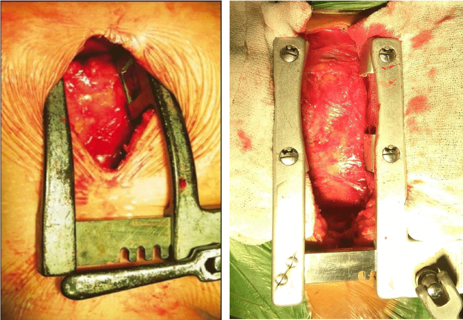
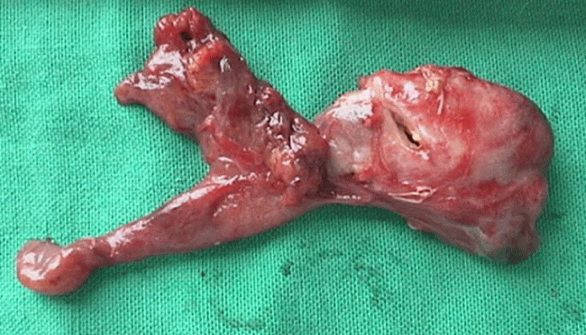
The patient is placed in a supine position with a pad under the shoulders. A vertical midline incision is made starting 2 cm below the suprasternal notch up to the level of the fourth intercostal space [10]. Longitudinal partial sternotomy provides adequate approach to the thymus. Advantage was, the incision may be inferiorly prolonged, by total sternotomy, or superiorly, by adding a transversal incision at the base of the neck. The incision is carried down through the subcutaneous tissue to expose the upper sternal border, presternal fascia, and the musculature, which are incised down to and through the sternal periosteum. The superior mediastinum, above the manubrium, is dissected with an electrocautery and bluntly with the finger to clear the posterior wall of the sternum away from the surrounding vascular structures. The bone of the manubrium is divided with a compressed air or an electric-powered saw in a downward dissection. The entire manubrium and the upper part of the sternal body down to the fourth intercostal space are divided (Figure 1). Subsequently, the upper mediastinum is exposed by Finochietto’s retractor with a lateral and progressive retraction. The thymus is then exposed. It must be totally removed, including its surrounding fat, starting superiorly from the base of the thyroid gland, using the phrenic nerves as lateral limits and proceeding inferiorly until the pericardium (Figure 2). Generally, we use a Duval clamp to expose the thymus and to allow sufficient tension for dissection of the thymus from the underlying structure. During dissection, the mediastinal pleura should be pushed laterally, to avoid inadvertently rupturing the pleura. If this should happen, it must be sutured immediately or ICD should be inserted at end of surgery. The surgeon should identify the phrenic nerves and avoid excessive manipulation and lesion, to avoid postoperative diaphragmatic palsy, which could seriously impair the clinical outcome of the patient. After resection, the thymic bed is carefully assessed to assure a radical thymectomy, adequate hemostasis, and that the pleural spaces are intact. A Portovac suction catheter is routinely placed close to the sternal notch. The sternum is then sutured with stainless steel wire no. 4 or 5, muscular layers are sutured with a 2-0 Vicryl thread, the subcutaneous and skin layers are sutured to conclude the procedure. A full sternotomy to complete the operation was not needed in our series. Successful management of these patients requires close cooperation among the neurologist, thoracic surgeon, anesthetist, nurses, and physiotherapists.
-

Figure 1:
Ministernotomy against full sternotomy.
-

Figure 2:
Excised thymectomy specimen.
The postoperative protocol consists of [Table 3]: (1) early extubation in the operating room; (2) 50% reduction in the administration of anticholinesterase drugs and removal of the mediastinal drain on the first postoperative day; (3) maintenance of the same dose of corticosteroid to the third postoperative week, and then reduce it to 5 mg/wk until a dose of 10 to 0 mg/d is obtained; and (4) immunosuppressive therapy with cytotoxic drugs and plasmapheresis is only used in patients with persistent symptoms or resistance to the use of corticosteroids. A neurological team is responsible for the late follow-up, therefore medication is controlled and adjusted according to the symptoms.
-

Table 3:
Postoperative Factors.
Results
There was no intraoperative or postoperative mortality. None of the patients required reoperation. In our comparitive study ministernotomy thymectomy has documented evidence of intraoperative as well postoperative early symptomatic recovery compared to full sternotomy thymectomy. Medication requirement decreased equally in both the groups. Ministernotomy technically simple, cosmeticaly better than full sternotomy with less pulmonary morbidity. We did not have any serious complications related to partial sternotomy; pain control is adequate with nonsteroidal analgesics, good cosmetic results, no reoperation in our series, no postoperative bleeding, no serious collections in the subcutaneous tissue, no diaphragmatic palsy were detected by roentgenogram, and no injury of the internal thoracic arteries.
Discussion
Although thymectomy has been accepted for the treatment of patients with MG, since the initial reports by Blalock and colleagues in 1939 [11], the surgical technique to be used, as well as clear indication of which patients should undergo surgical treatment, remain controversial and a matter of discussion. Many surgeons advocate minimal procedures to reduce morbidity and others are in favor of a more radical approach that provides better exposure, permitting total removal of the gland, but with greater morbidity and mortality. However, variations in anatomic configuration make it difficult or impossible to remove the total thymus tissue with most of these procedures. Because the striking effects of thymectomy on young adult patients with MG had been clarified, some investigators claimed that thymectomy should be performed in the elderly as well as in young patients. But few studies have dealt with the effect of the procedure on these populations. In 1985, Monden and colleagues [12] reported that in 27 elderly patients, the clinical effects of thymectomy were not statistically different from those in young adults. On the basis of those results, they believe that thymectomy should be performed in all elderly patients with MG. We believe that advanced age does not alter the outcome of thymectomy in patients with MG. Older adults can expect to have similar responses and require a similar number of postoperative medications as younger patients, but with a higher short-term morbidity and higher incidence of thymoma. Recently, two published articles confirmed our observations: Hamada and associates [13], in 1999, stated that although elderly patients are usually considered to be less responsive to an operation, thymectomy may sometimes be the treatment of choice for MG, even in octogenarians, and Evoli and co-workers [14], in 2001, also concluded that the prognosis of MG in older individuals seems to be favorable, although full remission is rare and MG weakness, treatment side effects, and associated thymoma can contribute to a higher mortality rate. Thymectomy may be carried out by median sternotomy [15, 16] (with or without a transversal cervical extension), partial sternotomy [17], a transcervical incision [5], transcervical incision combined to partial sternotomy [18, 19], and more recently, video-assisted thoracoscopy [8, 9, 20]. On the other hand, a more radical “maximal” thymectomy has been advocated by other investigators, such as Masaoka and colleagues [21], but this approach remains unpopular among surgeons performing thymectomy, as mentioned by Jaretzki and associates [22] in 1988.
As with any of these techniques, the surgeon must be extremely careful in resectioning the thymic tissue. If it is a cervical approach, total sternotomy, mini partial sternotomy, video thoracoscopy, or any other approach, the incision should be enlarged or the technique should be changed whenever there is the slightest chance of unsatisfactory resection. Removal should be deemed unsatisfactory whenever the thymus gland is torn by surgical traction and when the gland is withdrawn in pieces. At present, persistence of symptoms in patients with MG who have undergone previous thymectomy has been attributed to thymus remnants. Since 1983, Rosenberg and colleagues [23] reported that 85% of the thymic tissue was found in patients who underwent reoperation by trans-sternal approach and 67% of them improved clinically.
Scott and Detterbeck [24] reported on the total median sternotomy approach, opening both pleuras, in 100 consecutive patients. They reported good results, there was no mortality, and there was improvement or remission of MG in 78% of the patients. They believe that the advantage of the bilateral opening of the pleuras is to provide good visualization of the phrenic nerves, therefore decreasing the probability of damaging them and allowing the full resection of the fat adjacent to the thymus. We believe that systematic opening is not required, as it was corroborated by our results and by other researchers using the transcervical approach. On the other hand, the systematic opening of the pleuras may increase the incidence of postoperative pleural complications and certainly increases the pain resulting from the drainage of the pleural spaces. In the study by Grandjean and associates [17], pleural complications were reported in 6% of the patients. They also reported three cases of wound infection, and the pectoral muscle had to be used in one of them to repair sternal leakage. We believe that there is a great advantage to partial sternotomy, as the possibilities of wound infection, mediastinitis, and sternal destabilization are much lower.
Ferguson [25], reports on the transcervical approach to perform thymectomy. The advantage of this method involves only a relatively minor surgical procedure of short duration. The initial motivation for using this approach was its less invasiveness, that it requires a shorter hospital stay, and has a lower perioperative morbidity compared with the trans-sternal approach. In a literature review of 530 patients in four series, improvement with this approach was observed in 90% of the patients, 80% were asymptomatic and 51% had remission of the disease. We believe that this is a less invasive approach, with good aesthetic results and fast recovery according to literature data. However, it definitely provides poorer visualization of the thymus and surrounding structures, especially in cases of thymic hyperplasia, compared to total or partial longitudinal sternotomy.
More recently, the use of video-assisted thoracoscopy has become an additional therapeutical option for these patients. Yim and colleagues [26], have reported on the use of video-assisted thoracoscopy by approaching the right hemithorax. They performed thymectomy for MG in 22 patients. Conversion was required in 1 patient owing to intraoperative bleeding. When compared to total sternotomy, they reported aesthetic factors, shorter hospitalization time, and less pain as the major advantages of the procedure. However, they also concluded that: “the true role of this approach in thoracic surgery awaits long-term results, and even among the surgeons performing VATS thymectomy, there is controversy over the exact technique and, in particular, whether the thymus should be approached from the left or right.” We believe that video-assisted thoracoscopy is a good approach; however, when compared to partial sternotomy, it is more technically difficult and causes more pain. In women, the fact that incisions can be made near the inframammary sulcus is an advantage; however, it is minimized by the type and size of the incision performed. Ruckert and associates [27], carried out a comparative study of the pulmonary function after video-assisted thoracoscopy and total longitudinal sternotomy. They evaluated 10 patients randomized to each group and concluded that video-assisted thoracoscopy is better tolerated than total sternotomy. We believe that comparing video-assisted thoracoscopy to partial sternotomy may be a good study to perform. Recently, Grandjean and co-workers [17], reported on the use of partial sternotomy in 4 patients, using an inverted T based on one of the access sites used in minimally invasive heart procedures. The use of the T was not required in the 478 patients in our series, which results in a lower incidence of injury to the internal thoracic arteries. We also stated in our comments to the article [28], that this approach deserves the attention of all thoracic surgeons because it permits excellent visualization of the thymus gland, its vascular attachments, and all peripheral tissues in the mediastinal region. In addition, it is a simple technique to perform, easy to teach, uses conventional surgical instruments, thoracic residents spend only 50 minutes on each procedure, and reproducible results in different centers are possible. The comparison of the surgical results of thymectomy for the treatment of MG is difficult owing to the different protocols used, different times the procedure was carried out, and different preoperative and postoperative management among the different institutions. The study carried out by Nieto and colleagues [16], evaluating prognostic factors in MG treated by thymectomy in 61 patients approached in most of the cases by total longitudinal sternotomy, shows the great variability of the postoperative results obtained. They emphasize that the duration of the disease is a better indicator of postresection prognosis than gender or youth, concluding that the operation should be indicated early after the onset of symptoms. The results presented in this study are very similar to the literature data, regardless of the surgical technique used, showing a remission rate of 64% to 80%, and a rate of improvement of the clinical picture of 63% to 96%. The surgical approach by partial median sternotomy ensures easy and adequate approach to the gland, allowing a safe resection with adequate margins, removing all adjacent fat. This is important, as anatomic studies have evidenced the presence of thymic tissue in the mediastinal fat, which is usually not completely resected when the transcervical approach is used. Results were excellent: no operative mortality, significant improvement (complete or significant remission of symptoms) in 75.2% of the patients, and a partial improvement in 17.4% of the cases, according to Osserman’s classification. In view of the fundamental role of the thymus in the development of normal immunocompetent lymphocytes, it would seem advisable to be restrictive with thymectomy during the first year of life. After this period, however, the operation is clearly indicated in children with MG. As it was reported by Klingen and colleagues [29], since 1977, when in 11 of 12 children the clinical effect of the operation was very favorable, with uneventful postoperative course and the disease stabilized satisfactorily. Navarro Ahumada and associates [30], reported their experience in five children treated by transsternal radical thymectomy during a period of 5 years, and all of them were in complete remission after a mean postoperative period of 33 months. The final objective of the surgical treatment of the patient with MG is the long-term improvement of the clinical picture, regardless of the approach used.
Conclusion
Though VATS thymectomy given preference over ministernotomy thymectomy but there is no documented evidences available for same in literature and VATS facility is not available in all hospitals. The learning curve for the VATS is also high. In our comparision study ministernotomy thymectomy has documented evidence of intraopt as well postopt early symptomatic recovery compared to full sternotomy thymectomy with decreased medication requirement equal in both the groups. Thoracoscopic and Cervical thymectomy advantages are less postoperative morbidity, minimal discomfort, rapid functional recovery, shorter postoperative hospital stays. Limitations are facilities not available everywhere, technically difficult, risk of subtotal thymectomy and post thoracoscopy neuralgia. Ministernotomy approach will always maintain its place for thymectomy whatever newer approaches may develop. Taking this into consideration, and based on our experience, we conclude that the surgical treatment of MG by partial median sternotomy is adequate, with low morbidity and mortality, and therefore, very effective in the treatment of these patients.
-
-
- Heitmiller RF (1999) Myasthenia gravis: clinical features, pathogenesis, evaluation, and medical management. Semin Thorac Cardiovasc Surg 11: 41–46. Link: https://goo.gl/ZwX7Zq
- Drachman DB (1994) Myasthenia gravis. N Engl J Med 330: 1797–1810. Link: https://goo.gl/SGuKmR
- Grob D, Arsura EL, Brunner NG, Namba T (1987) The course of myasthenia gravis and therapies affecting outcome. Ann N Y Acad Sci 505: 472–499. Link: https://goo.gl/93MmeM
- Papatestas AE, Genkins G, Kornfeld P, Eisenkraft JB, Fagerstrom RP, et al. (1987) Effects of thymectomy in myasthenia gravis. Ann Surg 206: 79–88. Link: https://goo.gl/eAiiC3
- Kirschner PA, Osserman KE, Kark AE (1969) Studies in myasthenia gravis. Transcervical total thymectomy. JAMA 209: 906–910. Link: https://goo.gl/Wz9RnU
- Mulder DG, Herrmann C, Buckberg GD (1974) Effect of thymectomy in patients with myasthenia gravis. A sixteen year experience. Am J Surg 128: 202–206. Link: https://goo.gl/L9fxZP
- Detterbeck FC, Scott WW, Howard JF Jr, Egan TM, Keagy BA, et al. (1996) One hundred consecutive thymectomies for myasthenia gravis. Ann Thorac Surg 62: 242–245. Link: https://goo.gl/7BBwgp
- Yim AP, Kay RL, Ho JK (1995) Video-assisted thoracoscopic thymectomy for myasthenia gravis. Chest 108: 1440–1443. Link: https://goo.gl/3um7ta
- Mack MJ, Landreneau RJ, Yim AP, Hazelrigg SR, Scruggs GR (1996) Results of video-assisted thymectomy in patients with myasthenia gravis. J Thorac Cardiovasc Surg 112: 1352–1360. Link: https://goo.gl/miqwnu
- Nagre SW (2016) Hurdles for Starting Ministernotomy Aortic Valve Replacement Program. J Cardiovasc Med Cardiol 3: 035-037. Link: https://goo.gl/ddfqdp
- Osserman KE (1958) Myasthenia gravis. New York: Grune and Stratton Inc 79–86.
- Blalock A, Manson MF, Morgan HJ, Riven SS (1939) Myasthenia gravis and tumors of the thymic region: report of a case in which the tumor was removed. Ann Surg 110: 544–561. Link: https://goo.gl/qqTyNV
- Monden Y, Nakahara K, Fujii Y, Hashimoto J, Ohno K, et al. (1985) Myastenia gravis in elderly patients. Ann Thorac Surg 39:433–436. Link: https://goo.gl/uYfa53
- Hamada Y, Sakai Y, Ito H, Ichikawa H, Morishita Y (1999) Extended thymectomy for myasthenia gravis in an octagenarian. A case report. J Cardiovasc Surg (Torino) 40: 893–895. Link: https://goo.gl/JsSYU2
- Evoli A, Batocchi AP, Batocchi AP, Minisci C, Di Schino C, et al. (2000) Clinical characteristics and prognosis of myasthenia gravis in older people. J Am Geriatr Soc 48:1442–1448. Link: https://goo.gl/brcCsD
- Hatton PD, Diehl JT, Daly BD, Rheinlander HF, Johnson H, et al. (1989) Transsternal radical thymectomy for myasthenia gravis: a 15-year review. Ann Thorac Surg 47: 838–840. Link: https://goo.gl/yxiRtq
- Nieto IP, Robledo JP, Pajuelo MC, Montes JAR, Giron JG, et al. (1999) Prognostic factors for myasthenia gravis treated by thymectomy: review of 61 cases. Ann Thorac Surg 67: 1568–1571. Link: https://goo.gl/AxXvzU
- Grandjean JG, Lucchi M, Mariani MA (2000) Reversed-T upper mini-sternotomy for extended thymectomy in myasthenic patients. Ann Thorac Surg 70: 1423–1424. Link: https://goo.gl/xD7j9i
- Paletto AE, Maggi G (1982) Thymectomy in the treatment of myasthenia gravis: results in 320 patients. Int Surg 67: 13–16.
- LoCicero J 3rd (1996) The combined cervical and partial sternotomy approach for thymectomy. Chest Surg Clin N Am 6: 85–93. Link: https://goo.gl/6YsKnW
- Mineo TC, Pompeo E, Lerut TE, Bernardi G, Coosemans W, et al. (2000) Thoracoscopic thymectomy in autoimmune myasthenia: results of left-sided approach. Ann Thorac Surg 69: 1537–1541. Link: https://goo.gl/fKUmQB
- Masaoka A, Yamakawa Y, Niwa H, Fukai I, Kondo S, et al. (1996) Extended thymectomy for myasthenia gravis pacients: a 20-year review. Ann Thorac Surg 62: 853–859. Link: https://goo.gl/eHmreg
- Jaretzki A, Penn AS, Younger DS, Wolff M, Olarte MR, et al. (1988) “Maximal” thymectomy for myasthenia gravis: results. J Thorac Cardiovasc Surg 95: 747–757. Link: https://goo.gl/SW3NpK
- Rosenberg M, Jauregui WO, De Vega ME, Herrera MR, Roncoroni AJ (1983) Recurrence of thymic hyperplasia after thymectomy in myastenia gravis. Its importance as a cause of failure of surgical treatment. Am J Med 74: 78–82. Link: https://goo.gl/t93tsL
- Scott W, Detterbeck F (1999) Transsternal thymectomy for myasthenia gravis. Semin Thorac Cardiovasc Surg 11: 54–58. Link: https://goo.gl/w7w8hQ
- Ferguson MK (1999) Transcervical thymectomy. Semin Thorac Cardiovasc Surg 11: 59–64. Link: https://goo.gl/Cn6qnR
- Yim AP, Kay RL, Izzat MB, Ng SK (1999) Video-assisted thoracoscopic thymectomy for myasthenia gravis. Semin Thorac Cardiovasc Surg 11: 65–73. Link: https://goo.gl/5BpgB8
- Ruckert JC, Walter M, Muller JM (2000) Pulmonary function after thoracoscopic thymectomy versus median sternotomy for myasthenia gravis. Ann Thorac Surg 70: 1656–1661. Link: https://goo.gl/5EAbc3
- Campos JRM (2000) Invited commentary. Ann Thorac Surg 70: 1423–1425. Link: https://goo.gl/bTcWZv
- Klingen G, Johansson L, Westerholm CJ, Sundstroom C (1977) Transcervical thymectomy with the aid of mediastinoscopy for myastenia gravis: eight years’ experience. Ann Thorac Surg 23: 342–347. Link: https://goo.gl/vuHkbm


Table 1:
Patient Selection Criterias.
Criteria
Full Sternotomy
[15]
Mini Sternotomy
[15]
Male/Female
5/10
5/10
Age Group
20 To 30 years
20 to 30 years
Medicaly stabalised patients
15
15
CT evidence of thymoma
10
10