Journal of Cardiovascular Medicine and Cardiology
Characteristics of Coronary Artery Vulnerable Plaque on Coronary Computed Tomography Angiography (CCTA) and Optical Coherence Tomography (OCT) - A Review Article
Kishan Singh Rawat, Astha Kushwaha*, Tarvinder Bir Singh Buxi, Anurag Yadav, Samarjit Singh Ghuman, Arun Mohanty and Varun Holla
Department of Radiology, Sir Ganga Ram Hospital, Old Rajinder Nagar, New Delhi-110060, India
Cite this as
Rawat KS, Kushwaha A, Singh Buxi TB, Yadav A, Ghuman SS, Mohanty A, et al. Characteristics of Coronary Artery Vulnerable Plaque on Coronary Computed Tomography Angiography (CCTA) and Optical Coherence Tomography (OCT) - A Review Article. J Cardiovasc Med Cardiol. 2025;12(1):001-006. Available from: 10.17352/2455-2976.000218Copyright License
© 2025 Rawat KS, et al. This is an open-access article distributed under the terms of the Creative Commons Attribution License, which permits unrestricted use, distribution, and reproduction in any medium, provided the original author and source are credited.Background: The most common cause of sudden coronary deaths is atherosclerotic plaque rupture and subsequent coronary artery thrombosis. Plaques that have high risk features are called vulnerable plaques, and are the precursor of acute coronary events and are characterized by a large necrotic core and a thin fibrotic cap in histopathology. Coronary Computed Tomography Angiography (CCTA) is a clinically established tool for the diagnosis of coronary artery disease, evaluation of coronary artery stenosis and characterization of the plaque. OCT has emerged as the most accurate imaging modality for the evaluation of intracoronary Thin Cap Fibroatheroma (TCFA).
Methods: We performed a thorough online search of full text articles on characteristics of vulnerable plaque on CCTA and OCT in English literature. After doing a critical appraisal a comprehensive review has been described.
Conclusion: In this review we have described the features of vulnerable plaque like napkin ring sign, low attenuation index and positive remodeling index on CCTA with simultaneous evaluation of thin cap fibroatheroma on OCT.
Abbreviations
CCTA: Coronary Computed Tomography Angiography; TCFA: Thin Cap Fibroatheroma; IVUS: Intravascular Ultrasound; OCT: Optical Coherence Tomography; NIRS: Near-Infrared Spectroscopy; LAP: Low Attenuation Plaque; RI: Remodeling Index; MIP: Maximum Intensity Projection; VRT: Volume Rendering Technique
Introduction
Background
Cardiovascular disease continues to be the leading cause of death globally, despite advances in risk stratification, diagnostic tools and preventative therapies [1]. The etiological risk factors associated with cardiovascular diseases are well recognized and include hyperlipidemia, obesity, smoking, hypertension and, a sedentary lifestyle. These factors are responsible for more than 90% of the cardiovascular disease risks in all epidemiological studies [2].
Various invasive and non-invasive imaging modalities are available for characterization of vulnerable plaques. Invasive modalities like Intravascular Ultrasound (IVUS), Optical Coherence Tomography (OCT), Near-Infrared Spectroscopy (NIRS), Angioscopy, and Thermography provide the closest information to match the histopathology. However noninvasive imaging modalities like Coronary Computed Tomography Angiography (CCTA), Cardiovascular Magnetic Resonance Imaging (CMR), and Nuclear imaging are alternatives to the invasive procedure. Non-invasive imaging modalities are ideal for screening the vulnerable plaque and for evaluating its progression. CCTA evaluates both the coronary artery wall and the lumen in a quick, noninvasive, and relatively inexpensive way. CCTA helps in the assessment of coronary artery stenosis, the extent of coronary artery disease, and plaque morphology, and plays a significant role in the management of CAD to reduce the rate of cardiac events and cardiac mortality [3-5].
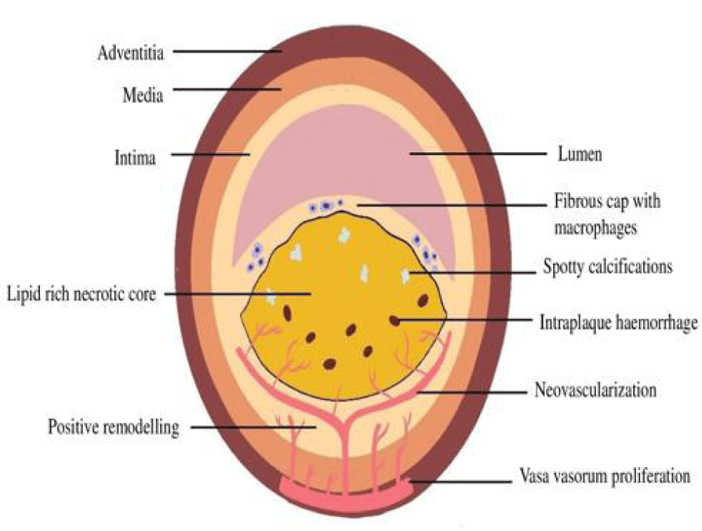
With newer imaging modalities like Optical Coherence Tomography (OCT) which is an optical analog of Intravascular Ultrasound (IVUS) coming into the picture, there has been a major interest in refining our current methods of diagnosis and individualizing preventative therapies like percutaneous coronary intervention. Myocardial infarction is most commonly caused by a rupture of atherosclerotic plaque. Plaques that are prone to rupture have certain common characteristics that together define the vulnerable plaque. There are hallmark characteristics of high-risk plaque that have been identified in histology (Figure 1), invasive intracoronary OCT and noninvasive Coronary Computed Tomography Angiography (CCTA) which serve as specific imaging modalities for coronary imaging. The Thin Cap Fibroatheroma (TCFA), a feature of vulnerable or high risk plaque is defined on OCT with findings comprising of fibrous cap of thickness < 65 µm overlying a lipid rich plaque with superficial microcalcification and plaque haemorrhage [6]. The vulnerable or high-risk plaque characteristics defined on CCTA include a Low Attenuation Plaque (LAP), Remodeling Index (RI), and napkin ring sign (NRS). It is worth mentioning that these findings are independent of stenosis severity and consistent with the fact that myocardial infarction is often due to plaque rupture in non-obstructive lesions [7].
In this article we review the role of CCTA in characterization of vulnerable plaque by assessing, vessel remodeling index (RI), Low Attenuation Plaque (LAP) and Napkin Ring Sign (NRS).
Coronary Computed Tomography Angiography (CCTA)
Clinically, CCTA has become the preferred noninvasive imaging technique for evaluating both lumen area and plaque composition in the majority of the coronary vasculature. This is especially true for vessels with a diameter greater than 1.5 mm, as this is where the majority of plaques are commonly situated [8]. Considerable benefits arise from its inherent characteristics, including its noninvasive nature, capacity to image the entire coronary vasculature, and the potential to evaluate both the vessel wall and lumen. It demonstrates outstanding capability in effectively ruling out conditions, with a negative prognostic value reaching nearly 100%, even during extended periods of follow-up [9,10].
The CCTA-derived parameters for high-risk plaques like positive vessel remodelling, low attenuation value of plaque, spotty calcification, and napkin ring sign have emerged as significant and independent predictors of events in multiple studies. The lack of invasiveness associated with CCCA makes it an appealing choice for population screening, particularly in the context of primary prevention. Its precision in identifying the presence of lesions is comparable to that of Intravascular Ultrasound (IVUS) [11].
Optical Coherence Tomography (OCT)
The precision of OCT imaging, with axial resolution ranging from 10 to 20 μm and lateral resolution from 20 to 40 μm, facilitates accurate observation of plaque morphology [12]. Additionally, it enables the assessment of fibrous cap thickness [12]. A distinctive capability of OCT includes the identification of macrophages, which typically range in size from 20 to 50 μm, as well as the detection of neovascularization and microcalcifications [13]. Limitations of OCT include the necessity for blood displacement due to the high scattering effect of its components, longer image acquisition times, frequent occurrence of artifacts, inability to assess deeper plaque structures, and relatively limited discrimination between calcified areas and lipid cores. This challenge arises because both present as signal-poor areas, with clear borders for calcified areas and diffuse borders for lipid cores.
Scan protocol for CT Coronary angiography (Table 1)
All patients should be in sinus rhythm at the time of study. For a patient with a heart rate (HR) > 60 beats per minute, steady HR of approximately 60 bpm should be achieved using beta blocker drugs and or an anxiolytic. Calcium channel blockers can be used in patients where beta blockers are contraindicated. Sublingual sorbitrate (2.5 mg) should be administered two minutes before scanning. After obtaining the initial scout images, a prospective gated ECG-triggered non-contrast scan is acquired for coronary calcium scoring. Retrospective gated ECG triggered contrast-enhanced cardiac CT angiography scan, with intravenous administration of a bolus of 80-110 ml of 350 mg/ml non ionic water-soluble contrast CONTRAPAQUE (Omnipaque: Iohexol 300) at a flow rate of 5 - 6 ml/s using 16 -18 G cannula followed by a saline flush of 20 ml, is acquired using 128 slice MDCT scanner.
Interpretation of CT Images
Curved multiplanar reformation, Maximum Intensity Projection (MIP), Volume Rendering Technique (VRT) images, 2-D and globe images were reconstructed and all scans were analyzed on a Philips workstation (Philips, Extended Brilliance Workspace and Intellispace portal software). Presence of a minimal three contiguous pixels with an attenuation of >130 H.U. was considered calcification.
The plaques were classified as calcified, non-calcified & mixed. Non-calified plaques were defined as structures assignable to the vessel wall (in at least two views) with a density lower than the lumen contrast whereas plaques demonstrating calcification less than or equal to 50% were classified as mixed. Similarly, plaques with more than or equal to 50% calcification were classified as calcified plaques.
The degree of stenosis was classified as mild (<50% stenosis, non-obstructive), moderate (50% - 75% stenosis), and severe (>75% stenosis). Greater than 50% stenosis was considered obstructive/significant CAD. For patients with varying degrees of stenosis in different coronary artery segments, the plaque with the highest degree of stenosis was used for the classification of degree of stenosis.
CT features of vulnerable plaque
There are two types of plaque: fibrocalcified plaque and vulnerable plaque. They both have a very different impact on the pathogenesis of the disease. The patients with fibrocalcified plaque present with stable angina however, those with vulnerable plaques are very prone to an acute cardiac event. It becomes very important to manage the precursor lesion of an acute cardiac event in an asymptomatic patient.
CT features of vulnerable plaque consist of a positive remodeling index, napkin ring sign, low attenuation plaque, and small luminal area.
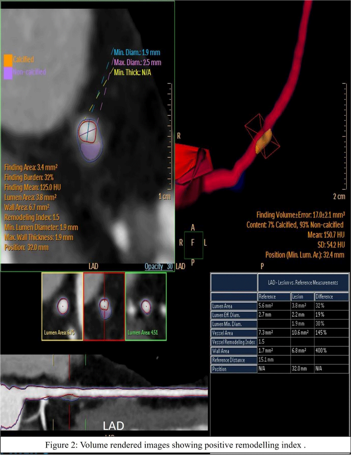
Remodelling index: The Remodeling Index (RI) (Figure 2) is calculated as the ratio of vessel diameter at the plaque site to a reference diameter of a normal-appearing vessel proximal to the lesion. Proximal and distal reference segments were defined as 5 mm long segments adjacent to the target segment. It was found in many studies, that plaques that have RI >1.1 are more commonly associated with vulnerable plaques [14].
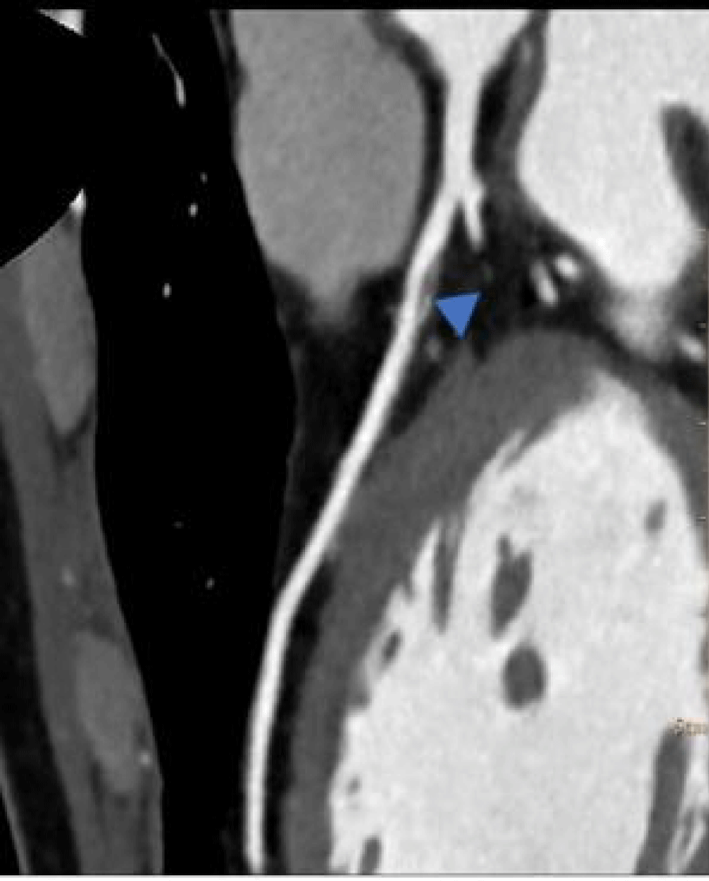
Napkin ring sign: Napkin Ring Sign (NRS) is defined as a semicircular thin rim enhancement around the center of plaque along the outer contour of the coronary artery, and the attenuation value of the rim should be more than the plaque but less than or equal to 130 Houndsfield unit (H.U.)(Figure 3)[15].
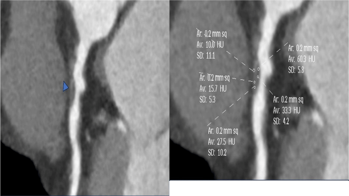
Low attenuation plaque: The attenuation value of plaque (in H.U.) was calculated as the average attenuation value of plaque after placing 5 rounded Regions of Interest (ROI) of 0.2 mm2 each. One ROI of 0.2 mm2 was also kept in the center of the lumen (Figure 4a,4b). Lowest H.U. in all ROI was measured & then, averaged. If the plaque was calcified then, ROI was placed in the non-calcified portion of the plaque. The plaques with attenuation value (HU>500) correspond with calcified plaques. Low Attenuation Plaque (LAP) was defined as a plaque with an attenuation value of <30 HU [16].
OCT protocol: OCT images are acquired using a nonocclusive technique with the C7XR system and DragonFly catheters (St Jude Medical, Minneapolis). The artery should be cleared of blood by continuous flushing with Iodixanol 370 at a flow rate of 3 ml/s for the right coronary artery and 4.5 ml/s for the left anterior descending and left circumflex. A 6 to 8 French size (0.014 inches) guide catheter is passed in the coronary artery under fluoroscopic guidance after administration of intra-coronary 100 - 200 microgram Nitroglycerine. Coronary guidewire is then, advanced proximal to the lesion & exchanged with an OCT imaging wire which is positioned distal to the lesion. OCT images were obtained with an automatic pullback of 20mm/s and the data was stored for offline analysis.
OCT features of vulnerable plaque: The study conducted by yabushita, et al. demonstrated that OCT has a high sensitivity (range 0.87 - 0.95) and specificity (range 0.94 - 1.0) for distinguishing between types of plaque [17]. On OCT, fibrous plaques show homogenous signal-rich layers and lipid-rich plaques were characterized by signal-poor sections with diffuse edges.
A vulnerable plaque on OCT is defined as a thin fibrous cap (<65 μm), a large lipid pool; and activated macrophages near the fibrous cap (Figure 5a,5b) [16].
Discussion
To reduce morbidity and mortality in suspected or proven coronary artery disease patients it is important to prevent acute coronary events. Both invasive and noninvasive modalities are there to identify vulnerable plaque. Noninvasive modality becomes ideal for screening asymptomatic patients and for assessing the disease progression. Due to its improved spatial and temporal resolutions, CCTA has the potential to quantify and characterize atherosclerotic plaques, particularly non-calcified vulnerable plaques.
According to pathological studies - plaque rupture, plaque erosion, and calcified nodules were three underlying pathologies leading to thrombosis of vessels and myocardial infarction. The thin cap fibroatheroma was identified as a prototypical high-risk plaque which is defined as a large, outwardly remodeled coronary lesion with a large necrotic core and a thin fibrous cap (<65 µm).
OCT is an invasive modality that can detect a TCFA plaque which is the feature of vulnerable or high-risk plaque.
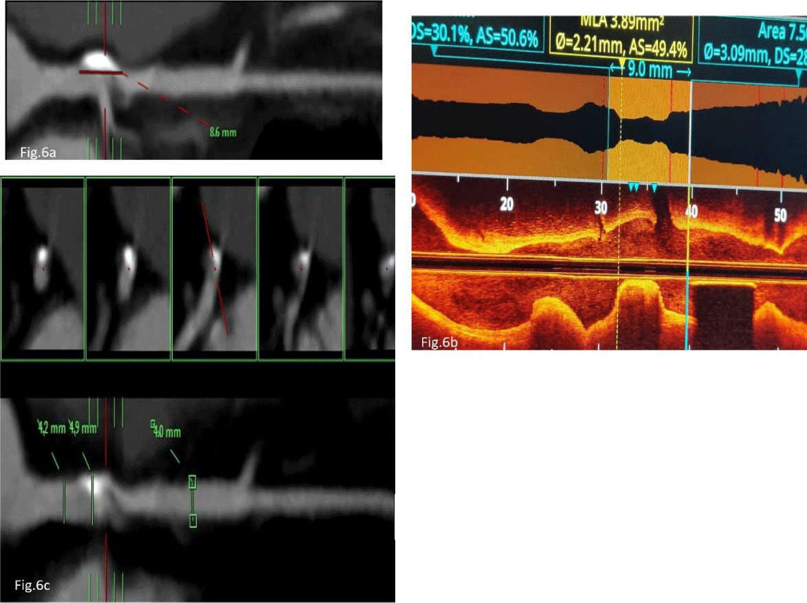
Many studies have shown that plaques that have high-risk features like NRS, (RI >1.1)(Figure 6) and LAP on CCTA are associated with TCFA on OCT.
This is in line with the study conducted by Yang et al who evaluated the presence of vulnerable plaque characteristics in per section to section-to-section study on CCTA was associated with the presence of TCFA as defined by optical coherence tomography (OCT) [18]. A total of 28 patients with 31 lesions were included in this study for analysis. In per-section analysis, OCT-TCFA showed higher frequency of CCTA-detected LAP (58.0% vs.18.5%), NRS (31.9% vs. 8.8%), and positive remodeling (68.1% vs. 48.0%) as compared to sections without OCT-TCFA (all p values were < 0.05).
The accuracy of CCTA in identifying thin-cap fibroatheroma (TCFA) detected by optical coherence tomography was studied by Tomizawa, et al. in 129 lesions in 106 patients [19]. They used three models to predict TCFA: Model 1 included R.I.>1; 1.1, minimum CT HU <30 HU and NRS; Model 2 included RI> 1.1, minimum CT HU <30 HU or NRS and last Model 3 included regression model using RI, proportion of LAP and NRS. They concluded that the accuracy to predict TCFA by CCTA would improve with a model using RI and LAP.
Nakazato et al and Kashiwagi, et al. also divided culprit plaques into TCFA and non-TCFA groups according to OCT criteria and compared the results with CCTA findings [20,21]. It was shown that TCFA plaques had lower attenuation values and NRS compared with non-TCFA plaques.
In a study conducted by Ozaki et al it was found that low attenuation plaque was seen in 88% of ruptured plaques and positive remodeling (RI >1.1) was seen in 96% of ruptured plaques [22]. Pflederer and Kashiwagi in their studies found that NRS had 96% to 100% specificity for the identification of TCFA [21,23]. It also aimed to correlate quantitative variables of coronary artery like plaque length, minimal lumen diameter, minimal lumen area, and diameter stenosis (%) between CCTA and OCT.
A study conducted by Horvart, et al. concluded that the assessment of the plaque characteristics improves the diagnostic accuracy of CCTA to identify advanced atherosclerotic lesions [15]. The CCTA finding of NRS has a high specificity and high positive predictive value for the presence of advanced lesions.
Dwivedi et al in a study reported that a plaque with low and intermediate attenuation (1 to 70 HU) is likely to be the most vulnerable, and a cutoff value for plaque volume of 3.99 mm/mm for combined low and intermediate plaque volume can be used as a strong prognostic indicator [24-26].
Conclusion
Imaging plays an important role in cardiovascular risk stratification. The identification and characterization of vulnerable plaques are critical in understanding and managing the risk of acute coronary events. Coronary Computed Tomography Angiography (CCTA) has proven to be a robust non-invasive tool, offering significant insights into plaque morphology, including features such as low attenuation plaque, positive remodeling index, and the napkin-ring sign. Simultaneously, Optical Coherence Tomography (OCT) provides unparalleled precision in identifying Thin-Cap Fibroatheromas (TCFA), the hallmark of high-risk plaques, at a microscopic level but is an invasive tool.
CCTA is a noninvasive technique for the detection of vulnerable plaque which can lead to an acute coronary event. CCTA not only helps in diagnosing but it plays a very important role in screening and follow-up of patients thus playing a pivotal role in primary and secondary prevention of acute coronary events. . Being a noninvasive technique it is becoming the choice of both clinicians and patients and sufficient data is there to prove its sensitivity and specificity.
- Hay SI, Abajobir AA, Abate KH, Abbafati C, Abbas KM, Abd-Allah F, et al. Global, regional, and national disability-adjusted life-years (DALYs) for 333 diseases and injuries and healthy life expectancy (HALE) for 195 countries and territories, 1990-2016: A systematic analysis for the Global Burden of Disease Study 2016. Lancet. 2017;390:1260-344. Available from: https://www.thelancet.com/journals/lancet/article/PIIS0140-6736(17)32130-X/fulltext
- Gagan D, Flora M, Nayak MK. A Brief Review of Cardiovascular Diseases, Associated Risk Factors and Current Treatment Regimes. Curr Pharm Des. 2019;25:4063-84. Available from: https://doi.org/10.2174/1381612825666190925163827
- Hamon M, Morello R, Riddell JW, Hamon M. Coronary arteries: diagnostic performance of 16- versus 64-section spiral CT compared with invasive coronary angiography—meta-analysis. Radiology. 2007;245:720-31. Available from: https://doi.org/10.1148/radiol.2453061899
- Motoyama S, Kondo T, Anno H, Sugiura A, Ito Y, Mori K, et al. Atherosclerotic plaque characterization by 0.5-mm-slice multislice computed tomographic imaging. Circ J. 2007;71:363-6. Available from: https://doi.org/10.1253/circj.71.363
- Motoyama S, Kondo T, Sarai M, Sugiura A, Harigaya H, Sato T, et al. Multislice computed tomographic characteristics of coronary lesions in acute coronary syndromes. J Am Coll Cardiol. 2007;50:319-26. Available from: https://doi.org/10.1016/j.jacc.2007.03.044
- Uemura S, Ishigami K, Soeda T, Okayama S, Sung JH, Nakagawa H, et al. Thin-cap fibroatheroma and microchannel findings in optical coherence tomography correlate with subsequent progression of coronary atheromatous plaques. Eur Heart J. 2012;33:78-85. Available from: https://doi.org/10.1093/eurheartj/ehr284
- Stone GW, Maehara A, Lansky AJ, de Bruyne B, Cristea E, Mintz GS, et al. PROSPECT Investigators. A prospective natural-history study of coronary atherosclerosis. N Engl J Med. 2011;364(3):226-35. Available from: https://doi.org/10.1056/nejmoa1002358
- Wang JC, Normand SL, Mauri L, Kuntz RE. Coronary artery spatial distribution of acute myocardial infarction occlusions. Circulation. 2004;110:278-84. Available from: https://doi.org/10.1161/01.cir.0000135468.67850.f4
- Andreini D, Pontone G, Mushtaq S, Bartorelli AL, Bertella E, Antonioli L, et al. A long-term prognostic value of coronary CT angiography in suspected coronary artery disease. JACC Cardiovasc Imaging. 2012;5:690-701. Available from: https://doi.org/10.1016/j.jcmg.2012.03.009
- Motoyama S, Ito H, Sarai M, Kondo T, Kawai H, Nagahara Y, et al. Plaque characterization by coronary computed tomography angiography and the likelihood of acute coronary events in mid-term follow-up. J Am Coll Cardiol. 2015;66:337-46. Available from: https://doi.org/10.1016/j.jacc.2015.05.069
- Voros S, Rinehart S, Qian Z, Joshi P, Vazquez G, Fischer C, et al. Coronary atherosclerosis imaging by coronary CT angiography: current status, correlation with intravascular interrogation and meta-analysis. JACC Cardiovasc Imaging. 2011;4:537-48. Available from: https://doi.org/10.1016/j.jcmg.2011.03.006
- Bom MJ, van der Heijden DJ, Kedhi E, van der Heyden J, Meuwissen M, Knaapen P, et al. Early Detection and Treatment of the Vulnerable Coronary Plaque: Can We Prevent Acute Coronary Syndromes? Circ Cardiovasc Imaging. 2017;10(5):e005973. Available from: https://doi.org/10.1161/circimaging.116.005973
- Tearney GJ, Regar E, Akasaka T, Adriaenssens T, Barlis P, Bezerra HG, et al. Consensus standards for acquisition, measurement, and reporting of intravascular optical coherence tomography studies: A report from the international working Group for Intravascular Optical Coherence Tomography Standardization and Validation. J Am Coll Cardiol. 2012;59(12):1058-1072. Available from: https://doi.org/10.1016/j.jacc.2011.09.079
- Glagov S, Weisenberg E, Zarins CK, Stankunavicius R, Kolettis GJ. Compensatory enlargement of human atherosclerotic coronary arteries. N Engl J Med. 1987;316:1371-1375. Available from: https://doi.org/10.1056/nejm198705283162204
- Maurovich-Horvat P, Schlett CL, Alkadhi H, Nakano M, Otsuka F, Stolzmann P, et al. The napkin-ring sign indicates advanced atherosclerotic lesions in coronary CT angiography. JACC Cardiovasc Imaging. 2012;5(12):1243-52. Available from: https://doi.org/10.1016/j.jcmg.2012.03.019
- Shmilovich H, Cheng VY, Tamarappoo BK, Dey D, Nakazato R, Gransar H, et al. Vulnerable plaque features on coronary CT angiography as markers of inducible regional myocardial hypoperfusion from severe coronary artery stenoses. Atherosclerosis. 2011;219:588-95. Available from: https://doi.org/10.1016/j.atherosclerosis.2011.07.128
- Yabushita H, Bouma BE, Houser SL, Aretz HT, Jang IK, Schlendorf KH, et al. Characterization of human atherosclerosis by optical coherence tomography. Circulation. 2002;106(13):1640-5. Available from: https://doi.org/10.1161/01.cir.0000029927.92825.f6
- Yang DH, Kang SJ, Koo HJ, Chang M, Kang JW, Lim TH, et al. Coronary CT angiography characteristics of OCT-defined thin-cap fibroatheroma: a section-to-section comparison study. Eur Radiol. 2018;28:833-43. Available from: https://doi.org/10.1007/s00330-017-4992-8
- Tomizawa N, Yamamoto K, Inoh S, Nojo T, Nakamura S. Accuracy of computed tomography angiography to identify thin-cap fibroatheroma detected by optical coherence tomography. J Cardiovasc Comput Tomogr. 2017;11(2):129-134. Available from: https://doi.org/10.1016/j.jcct.2017.01.010
- Nakazato R, Otake H, Konishi A, Iwasaki M, Koo BK, Fukuya H, et al. Atherosclerotic plaque characterization by CT angiography for identification of high-risk coronary artery lesions: A comparison to optical coherence tomography. Eur Heart J Cardiovasc Imaging. 2015;16:373-9. Available from: https://doi.org/10.1093/ehjci/jeu188
- Kashiwagi M, Tanaka A, Kitabata H, Tsujioka H, Kataiwa H, Komukai K, et al. Feasibility of noninvasive assessment of thin-cap fibroatheroma by multidetector computed tomography. JACC Cardiovasc Imaging. 2009;2:1412-1419. Available from: https://doi.org/10.1016/j.jcmg.2009.09.012
- Ozaki Y, Okumura M, Ismail TF, Motoyama S, Naruse H, Hattori K, et al. Coronary CT angiographic characteristics of culprit lesions in acute coronary syndromes not related to plaque rupture as defined by optical coherence tomography and angioscopy. Eur Heart J. 2011;32:2814-2823. Available from: https://doi.org/10.1093/eurheartj/ehr189
- Maurovich-Horvat P, Hoffmann U, Vorpahl M, Nakano M, Virmani R, Alkadhi H. The napkin-ring sign: CT signature of high risk coronary plaques? JACC Cardiovasc Imaging. 2010;3:440-444. Available from: https://doi.org/10.1016/j.jcmg.2010.02.003
- Pflederer T, Marwan M, Schepis T, Ropers D, Seltmann M, Muschiol G, et al. Characterization of culprit lesions in acute coronary syndromes using coronary dual-source CT angiography. Atherosclerosis. 2010;211:437-444. Available from: https://doi.org/10.1016/j.atherosclerosis.2010.02.001
- Dwivedi G, Liu Y, Tewari S, Inacio J, Pelletier-Galarneau M, Chow BJW. Incremental Prognostic Value of Quantified Vulnerable Plaque by Cardiac Computed Tomography. J Thorac Imaging. 2016;31:373-379. Available from: https://doi.org/10.1097/rti.0000000000000236
- Glagov S, Weisenberg E, Zarins CK, Stankunavicius R, Kolettis GJ. Compensatory enlargement of human atherosclerotic coronary arteries. N Engl J Med. 1987;316:1371-1375. Available from: https://doi.org/10.1056/nejm198705283162204
