Journal of Cardiovascular Medicine and Cardiology
Prevalence of valvar regurgitation in Nigerian children with structurally normal hearts using colour doppler echocardiography
Chika O Duru1*, Josephat M Chinawa2, Igoche D Peter3, Ibrahim Aliyu4, Mustafa O Asani4 and Fidelia Bode-Thomas5
2Department of Pediatrics, University of Nigeria Teaching Hospital, Ituku-Ozalla, Enugu, Nigeria
3Division of Pediatrics Cardiology, The Limi Children’s Hospital Abuja, Nigeria
4Department of Pediatrics Aminu Kano Teaching Hospital, Kano, Nigeria
5Department of Pediatrics, University of Jos Teaching Hospital, Jos, Plateau State, Nigeria
Cite this as
Duru CO, Chinawa JM, Peter ID, Aliyu I, Asani MO, et al. (2020) Prevalence of valvar regurgitation in Nigerian children with structurally normal hearts using colour doppler echocardiography. J Cardiovasc Med Cardiol 7(3): 262-267. DOI: 10.17352/2455-2976.000149Background: Doppler echocardiography is a reliable and non-invasive method of detecting valvar regurgitation. The aim of this study was to determine the prevalence of valvar regurgitation in children with structurally normal hearts and explore its relationship with age, gender and anthropometry.
Methods: This was a descriptive cross-sectional study conducted in four tertiary hospitals in Nigeria. Two hundred and thirty-three children (124 males and 109 females), from birth to 18 years were recruited prospectively. Using transthoracic echocardiography, the presence of valvar regurgitation was assessed across the four cardiac valves using Color Doppler interrogation after structural abnormalities were ruled out. Data was analyzed using the Statistical Package for Social Sciences (SPSS) software, version 22.
Results: Valvar regurgitation was found in 158/233 children giving a prevalence of 67.8%. They consisted of 87 males and 71 females with a male: female ratio of 1.2:1. Of these, 82 (35.2%) had a single valvar regurgitation, 72 (30.9%) had regurgitation at two valves while 4 (1.7%) had regurgitation at three valves. Pulmonary regurgitation was the most common in 52.8% of cases while aortic regurgitation was seen in only two children (0.9%). There was a non-significant negative correlation of age and body surface area with presence of tricuspid regurgitation (ρ -0.79, p=0.22 and ρ -0.12, p=0.08) and mitral regurgitation (ρ -0.04, p=0.56 and ρ -0.02, p=0.83) but positive correlation with pulmonary regurgitation (ρ 0.11, p= 0.11 and ρ 0.12, p= 0.08). The presence of valvar regurgitation was not associated with gender (p=.>0.05).
Conclusion: The prevalence of valvar regurgitation in apparently healthy Nigerian children is 67.8%. The presence of valvar regurgitation especially of the right sided valves could be a physiologic finding in apparently healthy children with structurally normal hearts. Regurgitation of the left sided valves is rare.
Abbreviations
MR: Mitral Regurgitation; TR: Tricuspid Regurgitation; PR: Pulmonary Regurgitation; AR: Aortic Regurgitation
Background
Transthoracic echocardiography with Doppler interrogation is a safe and reliable way of detecting valvar regurgitation [1-6]. Physiologic valvar regurgitation is usually suspected during echocardiographic evaluation of children with chest pain or palpitations who may not have underlying congenital heart disease. On auscultation of the cardiovascular system, however valvar regurgitation can present with murmurs or thrills which occur due to the turbulence of blood flowing through the valve orifices [4,7]. Insignificant valvar regurgitations are considered physiological in clinical practice and can easily be missed on clinical examination [1-3,6,7]. Doppler echocardiography provides a means of assessing the presence, etiology and severity of the regurgitation and effect of its presence on cardiac size and function [4-7].
Several studies have described the prevalence of valvar regurgitation in adults and children with structurally normal hearts [3, 8-12]. Regurgitation of the pulmonary and tricuspid valves which are the right sided heart valves have been widely reported by several authors [10-12]. In addition to the predominance of these right sided heart regurgitant lesions, their prevalence has been noted to increase with advancing age [12,13]. The presence of regurgitation of the left sided heart valves especially the aortic valve usually indicates a pathological process and thus is rarely seen in normal hearts [6-9]. It is thus imperative to have a baseline information on the normal valvar findings in structurally normal hearts. There have been no previous studies on prevalence of valvar regurgitation in apparently healthy children or adults in Nigeria. Thus the aim of this study was to determine the prevalence of valvar regurgitation in Nigerian children with structurally normal hearts and explore its relationship with age, gender and anthropometry.
Methods
Study centers
This was a cross sectional descriptive multicenter study carried out at the Paediatric cardiology clinics of the Niger Delta University Teaching Hospital (NDUTH) Okolobiri, University of Nigeria Teaching Hospital (UNTH), Ituku-Ozalla, Enugu and Aminu Kano Teaching Hospital (AKTH) Kano. The three hospitals are tertiary centers located in South-south, South-East and North-Western parts of Nigeria with active paediatric cardiology practices.
Sampling
A total of two hundred and thirty-three (233) apparently healthy children from the three centers who were aged between 1 day and 18 years were recruited by convenience sampling methods after written informed consent. Each participant had a thorough physical examination and anthropometry performed. Weight was measured in kilograms using a SECA® weighing scale to the nearest 0.1kg. Height was measured in centimeters with the child placed on a fixed stadiometer to the nearest 0.1cm and converted to meters. Body surface area was derived for each participant using the Mosteller formula [14], Thereafter, in accordance with recommendations by the American Society of Echocardiography [15], a transthoracic echocardiogram, was performed on each child by experienced paediatric cardiologists in the respective institutions. A portable My Lab Gamma Esaote® cardiac ultrasound machine was used at NDUTH, E2 Sonoscape cardiac ultrasound machine at UNTH while SSI-8000 Sonoscape cardiac ultrasound machine was used at AKTH. Each machine was fitted with appropriate frequency probes depending on the age of the child and image quality. After ruling out structural abnormalities using 2-dimensional echocardiography, the four valve areas (mitral, tricuspid, aortic and pulmonary) were carefully examined for the presence of any malformed or thickened leaflets. When the leaflets were ascertained to be morphologically normal, each valve was interrogated using color Doppler imaging to detect the presence of regurgitant jets.
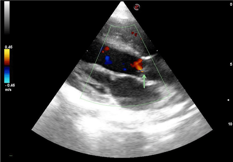
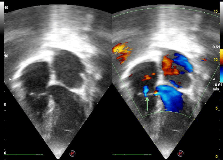
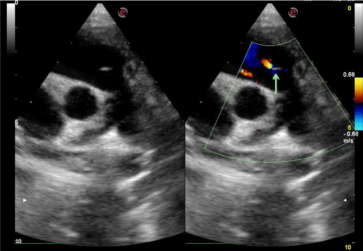
Doppler windows were the parasternal long-axis, parasternal short-axis, and apical five-chamber views for the aortic valve (Figure 1); and the parasternal long-axis, parasternal short-axis, and apical four-chamber views for the mitral valve. For the tricuspid valve, the right ventricular inflow view and parasternal and apical four and five chamber views were used (Figure 2) while the right ventricular outflow tract and the high parasternal short-axis view was used for the pulmonary valve (Figure 3). The flow signal was adjudged as regurgitant when it was observed as a reversed flow away from the valve by color Doppler echocardiography and when it lasted more than 100 msec by M-mode color flow mapping [6,12].
Study subjects
The study subjects were apparently healthy children between the ages of 1 day and 18 years of age who were recruited consecutively from the paediatric outpatient clinics of the hospitals during well child or follow up visits. Those who were above the ages of 18 years and who had known underlying cardiac disease (congenital or acquired) or any other chronic illness (HIV, tuberculosis, renal disease, diabetes or sickle cell anemia) or whose parents refused consent for the study were excluded.
Ethical considerations
Ethical approval was obtained from the Research and Ethics Committees of the three participating hospitals. Written informed consent was obtained from the parents of the children and assent from children aged 7 years and above.
Statistical analysis
Data was analyzed using the Statistical Package for Social Sciences (SPSS) software, version 22 (IBM, Armonk, NY, USA). Categorical data were described as frequencies and percentages. Continuous variables were assessed for normality using the Kolmogorov-Smirnov test. Descriptive statistics including mean ± standard deviation and median with (interquartile range) were used to summarize continuous data that were normally or non-normally distributed respectively. Chi square was used to determine the association between the presence of valvar regurgitation and gender. Correlation of the various valvar regurgitations (mitral, tricuspid and pulmonary) with BSA and age were examined using Spearman Rank order correlation (as these data were all non-normally distributed). A p- value < 0.05 was considered statistically significant.
Results
A total of 233 apparently healthy subjects were recruited for this study. There were 124 males and 109 females, with a male: female ratio of 1.1:1 The participants were aged between 1 day and 18 years with a median age of 98 months (IQR 60.0-143.4). They had a median weight of 8.25 kg (IQR 2.0-29.0), mean height of 124.6 cm (±28.8), and a median body surface area of 0.92 m2 (IQR 0.71-1.2).
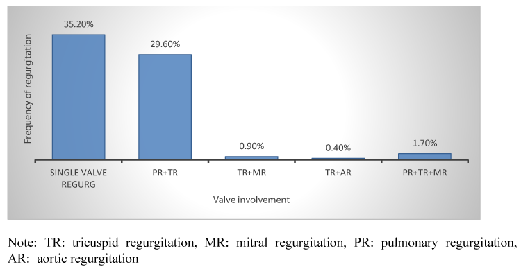
One hundred and fifty-eight; 158 children had at least one regurgitant valve giving the prevalence of valvar regurgitation to be 67.8%. They consisted of 87 males and 71 females with a male: female ratio of 1.2:1. Of these, 82 (35.2%) had a single valvar regurgitation, 72 (30.9%) had regurgitation of two valves while 4 (1.7%) had regurgitation at three valves.
Of the 82 with single valve regurgitation, only pulmonary regurgitation (PR) and tricuspid regurgitation (TR) were observed in 85.7% and 14.3% of the valves respectively.
Of the 72 with regurgitation noted in two valves, majority; 69 (95.8%) had PR co-existing with TR, 2 (2.8%) had TR with mitral regurgitation (MR) while 1 (1.4%) had TR with aortic regurgitation (AR) – these constituted 29.6%, 0.9% and 0.4% of the overall valvar regurgitations respectively. Only 4 children had regurgitation across three valves (PR+TR+MR) (Figure 4).
In terms of the frequency of regurgitation at each valve, pulmonary valvar regurgitation was the most common while aortic valvar regurgitation was the least common with frequencies of 52.8% and 0.9% respectively (Table 1). Of the 123 (52.8%) subjects with PR, males out-numbered the females (71 vs 52). Similarly, tricuspid regurgitation in 107 (45.9%) subjects was commoner in males (63) compared with females (44), while an equal number of boys and girls had MR (3 each) and only 2 boys had AR.
Though there was a male preponderance of cases with valvar regurgitation, there was no statistically significant association between gender and presence of TR (p=0.07), MR (p=1.00) and PR (p=0.09) (Table 2). There was a negative correlation of age with presence of TR (ρ -0.79, p=0.22) and MR (ρ -0.04, p=0.56) but positive correlation with PR (ρ 0.11, p= 0.11) though these were not statistically significant. Similarly, there was a negative correlation of body surface area with presence of TR (ρ -0.12, p=0.08) and MR (ρ -0.02, p=0.83) but positive correlation with PR (ρ 0.12, p 0.08) were was also not statistically significant (Table 3).
Discussion
The prevalence of valvar regurgitation in our study was 67.8%. This figure was similar to the 59.7% reported by Ayabakan, et al. [10], whose study in Turkish children had a similar age distribution. Our finding was also similar to the prevalence of 61.4% as reported by Lee, et al. [11], in the assessment of 420 infants in Taiwan. Yoshida, et al. [6] whose study consisted of subjects from 6 to 49 years reported prevalence of 45-85% among the varying age groups. However, Brand, et al. [3] and Choong, et al. [13], reported much lower prevalence rates of 26.9% and 34.0% of valvar regurgitation among children in their studies. Schmidt, et al. [5], noted that variations in Doppler echocardiographic methods for quantifying valvar regurgitation which could account for these differences. They could also be due to variations in sample size, race and study methodology.
In our study, single valvar regurgitation and regurgitation at two valves were seen in 35.2% and 30.9% respectively with regurgitation at three valves occurring in less than 2% of cases. Single valvar regurgitation has been noted to be more prevalent than multiple valvar regurgitation by other authors [3,11]. It was also noted that these single valvar regurgitations only involved the right sided valves which we also found to show more regurgitation than the left sided valves. Right valvar regurgitation predominance in normal hearts has similarly been described in previous studies although with variations in individual valvar regurgitation [3,6,10-12,16]. The prevalence of pulmonary regurgitation (PR) noted in our study was slightly higher than that of the tricuspid regurgitation (TR) with prevalence of 52.8% and 45.9% respectively. Van Diyk, et al. [12], reported a much higher prevalence of PR (84.0%) compared to TR (8.0%) in 173 children between the ages of 5 and 14 years in their study. Right sided valvar regurgitation has been noted to be common in children and adults and is usually physiologic [4,6,17]. Some authors have suggested that the frequency of right-sided regurgitation may be related to the closer positioning of the transducer to the right side of the heart which reduces interferences and enhances image quality of Doppler signals [10]. The low prevalence of left sided valvar regurgitation in our study has also been reported by other authors [9,16,17]. Thompson, et al. [9], reported a prevalence of <2% of MR among 32 children in their study and AR in only one child. As shown in ours and previous studies, AR is rare in children and adults and when detected, should be regarded as pathologic [6,17,18]. The higher left heart pressure compared with the right, higher systemic vascular resistance and intimal proliferation of the left heart chamber vasculatures may also explain the low prevalence of left heart regurgitation seen in this and other studies [19].
Though there was a male preponderance of subjects with right sided regurgitation (PR and TR), this difference was not statistically significant. Ayabakan, et al. [10], found that TR was more prevalent in the male subjects but noted no association of the other valvar regurgitation with gender. Gender differences in valvar heart diseases have been described in several studies with females having more mitral valve regurgitation than males [20-24].However, this has been observed mainly in females with organic valvar heart lesions such as degenerative and rheumatic mitral valve diseases [20-22] whereas our study involved only normal valves. Only two of our subjects had AR and both were male. Though the prevalence of AR has been reported to be low in subjects less than 60 years [17], it has been found to be commoner in males, though this may be due to the fact that most of these studies involved more male subjects than females [18,21]. Tricuspid regurgitation has been reported to be predominant in females in some studies and has been attributed to annular dilatation from chronic pressure and volume overload [8,25].
There was a negative correlation of age and body surface area with the presence of TR and MR which implied that the regurgitation of these valves are less prevalent with increasing age and body surface area. The presence of physiological MR has been noted to be commoner in the female gender [17,24]. Jones, et al. [24], noted an increase in the presence and severity of MR in those with lower BMI, female gender, mitral valve prolapse and stenosis and older ages in 3486 American Indian subjects. Our study also noted a positive correlation of age and body surface area with PR which implies that the prevalence of PR increases with age and body surface area though these relationships were not statistically significant. However Brand, et al. [3], noted that the prevalence of PR increased significantly with age unlike the regurgitations noted in the other valves [3]. Van Diyk, et al. [12], reported that neither age nor gender had any effect on the prevalence of right sided valvar regurgitation in their study. Choong, et al. [13], however noted an increased prevalence of MR, TR and AR with increasing age unlike findings from Yoshida, et al. [6]who studied 211 subjects from the ages of 6 to 49 years and noted a decreasing prevalence of TR, PR and MR with age. Okura, et al. [17], however reported that TR was the most prevalent regurgitant jet in >80% of subjects between 10 and 89 years in their study and this prevalence was similar across the various age groups. The differences noted in these various studies could be attributed to differences in methodology and sampling. However, the increased prevalence of valvar regurgitation in young subjects could be attributed to the better ultrasonic penetration which is due to their fattened bony thorax, leaner body mass and lower body fat which permits adequate detection of Doppler signals [6,26].
This study does have some limitations. It was a qualitative assessment of valvar regurgitation parameters and did not involve measurements of velocities and other functional parameters. This however is recommended to be explored in further studies.
Conclusion
The prevalence of valvar regurgitation in apparently healthy Nigerian children is 67.8%. Regurgitation of the right sided valves is commoner than left sided valvar regurgitation with PR being the most prevalent. Regurgitation at the aortic valve is very rare. Presence of valvar regurgitation especially of the right sided valves could be a physiologic finding in apparently healthy children with structurally normal hearts.
Ethical approval and consent to participate
Ethical approval was obtained from the Research and Ethics Committees of the Niger Delta University Teaching Hospital Okolobiri Bayelsa State Nigeria (NDUTH/REC/2020/0008) and the University of Nigeria Teaching Hospital, Ituku-Ozalla, Enugu State, Nigeria (NHRED/05/01/2008B-FWA00002458-1RB00002323) and Aminu Kanu Teaching Hospital Kano (NHREC/21/08/2008/AKTH/EC/1296). Written informed consent was obtained from the parents of the study participants before conduct of the study.
Funding
This study was self-funded. The authors did not receive any support from a grant or any other funding body.
Authors contributions
COD conceived the study and wrote the first draft of the manuscript. JMC and IDP collected, analyzed and interpreted the data. IA, MOA and FB critically reviewed the manuscript. All the authors approved of the final version to be published.
We gratefully acknowledge all the participants of the study.
- Richards KL, Cannon SR, Crawford MH, Sorensen SG (1983) Noninvasive diagnosis of aortic regurgitation and mitral valve disease with pulsed Doppler spectral analysis. Am J Cardiol 51: 1122-11277. Link: https://bit.ly/3259ujq
- Rahko PS (1989) Prevalence of Regurgitant Murmurs in Patients with Valvular Regurgitation detected by Doppler Echocardiography. Ann Intern Med 111: 466-472. Link: https://bit.ly/2EbHBhS
- Brand A, Dollberg S, Keren A (1992) The prevalence of valvular regurgitation in children with structurally normal hearts: a color Doppler echocardiographic study. Am Heart J 123: 177-80. Link: https://bit.ly/3l90bYO
- Zoghbi WA, Adams D, Bonow RO, Enriquez-Sarano M, Foster E, et al. (2017) Recommendations for the non-invasive evaluation of native valvular regurgitation. A report from the American Society of Echocardiography. Developed in collaboration with the Society for Cardiovascular Magnetic Resonance. J Am Soc Echocardiogr 30: 303-371. Link: https://bit.ly/3l3jAKs
- Schmidt A, Almeida OC, Pazin A, Marin-Neto JA, Maciel BC (2000) Valvular Regurgitation by Color Doppler Echocardiography. Arq Bras Cardiol 74: 273-281. Link: https://bit.ly/31ejibG
- Yoshida K, Yoshikawa J, Shakudo M, Akasaka T, Jyo Y, et al. (1988) Color Doppler Evaluation of Valvular Regurgitation in Normal Subjects. Circulation 78: 840-847. Link: https://bit.ly/3hkyJEQ
- Sahn DJ, Maciel BC (1988) Physiological valvular regurgitation. Doppler echocardiography and the potential for iatrogenic heart disease. Circulation 78: 1075 – 1077. Link: https://bit.ly/3aIEREw
- Singh JP, Evans JC, Levy D, Larson MG, Freed LA, Fuller DL, et al. (1999) Prevalence and clinical determinants of mitral, tricuspid, and aortic regurgitation (the Framingham heart study). J Am Coll Cardiol 83: 897-902. Link: https://bit.ly/2FJhTSm
- Thompson JDR, Allen J, Cribbs JL (2000) Left sided valvar regurgitation in normal children and adolescents. Heart 83: 185-187. Link: https://bit.ly/2FKwxbZ
- Ayabakan C, Özkutlu S, Kýlýç A (2003) The Doppler echocardiographic assessment of valvular regurgitation in normal children. Turk J Pediatr 45: 102-107. Link: https://bit.ly/2ErWGLO
- Lee ST, Lin MT (2010) Color Doppler Echocardiographic assessment of valvar regurgitation in normal infants. J Formos Med Assoc 109: 56-61. Link: https://bit.ly/2YowLMu
- Van Dijk APJ, Van Oort AM, Daniels O (1994) Right –sided valvular regurgitation in normal children determined by combined colour-coded and continuous wave Doppler echocardiography. Acts Paediatr 83: 200-203. Link: https://bit.ly/3aH0Vzt
- Choong CY, Abascal VM, Weyman J, Levine RA, Gentile F, et al. (1989) prevalence of valvular regurgitation by Doppler echocardiography in patients with structurally normal hearts by two-dimensional echocardiography. Am Heart J 117: 636-642. Link: https://bit.ly/3aGHCpW
- Mostellar RD (1987) Simplified calculation of body surface area. N Engl J Med 317: 1098. Link: https://bit.ly/3l6Dynt
- Lai WW, Geva T, Shirali GS, Frommelt PC, Humes RA, et al. (2006) Guidelines and standards for performance of a pediatric echocardiogram: A Report for the Task Force of the Pediatric Council of the American Society of Echocardiography. J Am Soc Echocardiogr 19: 1413-1430. Link: https://bit.ly/2Ej96FY
- Da Silva Mattos S, Severi R, Cavalcanti CV, Da Fonte Freire M, et al. (1992) Valvar regurgitation in normal children, Is it clinically significant? Cardiology in the Young 2: 291-297. Link: https://bit.ly/2E3ju53
- Okura H, Takada Y, Yamabe A, Ozaki T, Yamagishi H, et al. (2011) Prevalence and Correlates of Physiological Valvular Regurgitation in Healthy Subjects. A Color Doppler Echocardiographic Study in the Current Era. Circ J 75: 2699-2704. Link: https://bit.ly/2YkC27K
- Lebowitz NE, Bella JN, Roman MJ, Liu JE, Fishman DP, et al. (2000) Prevalence and correlates of aortic regurgitation in American Indians: the Strong Heart Study. J Am Coll Cardiol 36: 461–467. Link: https://bit.ly/2Q8TDee
- Avierinos JF, Inamo J, Grigioni F, Gersh B, Shub C, et al. (2008) Sex differences in morphology and outcomes of mitral valve prolapse. Ann Intern Med 149: 787-795. Link: https://bit.ly/2Q9wyIo
- Nitsche C, Koschutnik M, Kammerlander A, Hengstenberg C, Mascherbauer J (2020) Gender-specific differences in valvular heart disease. Wien Klin Wochenschr 132: 61-68. Link: https://bit.ly/2FCLsos
- Tornos P, Sambola A, Permanyer-Miralda G, Evangelista A, Gomez Z, et al. (2006) Long-term outcome of surgically treated aortic regurgitation: influence of guideline adherence toward early surgery. J Am Coll Cardiol 47: 1012-1017. Link: https://bit.ly/34kSGHW
- Marsan NA (2019) Gender difference in mitral valve disease: Where is the bias? Eur J Prev Cardiol 26: 1430-1432. Link: https://bit.ly/3aHP18x
- Shi Y, Wan Y, Wang Y, Wang K, Wang Q (2017) Mechanism underlying gender difference in heart disease risks and corresponding preventive measures. Eur J Prev Cardiol 24: 1807-1808. Link: https://bit.ly/2EpcajK
- Jones EC, Devereux RB, Roman MJ, Liu JE, Fishman D, et al. (2001) Prevalence and correlates of mitral regurgitation in a population-based sample (the Strong Heart Study). Am J Cardiol 87: 298-304. Link: https://bit.ly/3l6E3hl
- Arsalan M, Walther T, Smith RL, Grayburn PA (2017) Tricuspid regurgitation diagnosis and treatment. Eur Heart J 38: 634-638. Link: https://bit.ly/32eLM4j
- Cebeci BS, Kardesoglu E, Celik T, Demiralp E (2004) Echocardiographical Characteristics of Healthy Young Subjects with Physiological Mitral Regurgitation. Journal of International Medical Research 32: 240-244. Link: https://bit.ly/3l6PyWn


