Journal of Cardiovascular Medicine and Cardiology
Association of central blood pressure profile, ambulatory blood pressure parameters with 2D echocardiographic diastolic dysfunction and carotid intima-media thickness in patients with coronary artery disease
Arulkumar Jegavanthan*, Lakshman Bandara, Thilina Jayasekara, Hansa Sooriyagoda and Thangarajah Jeyakanth
Cite this as
Jegavanthan A, Bandara L, Jayasekara T, Sooriyagoda H, Jeyakanth T (2021) Association of central blood pressure profile, ambulatory blood pressure parameters with 2D echocardiographic diastolic dysfunction and carotid intima-media thickness in patients with coronary artery disease. J Cardiovasc Med Cardiol 8(1): 001-006. DOI: 10.17352/2455-2976.000159Introduction: Blood Pressure (BP) and its diurnal variability are linked to endothelial dysfunction and arterial stiffness, promoting atherosclerosis which can be observed as intimal thickening in carotid arteries and atherosclerotic lesions in coronary angiograms.
Objectives: To evaluate the association of Diastolic Dysfunction (DD) in patients with Coronary Artery Disease (CAD) with central and Ambulatory Blood Pressure Monitoring (ABPM) indices and to assess Carotid Intima Media Thickness (CIMT) in different CAD score groups and then to compare the results with individuals having normal coronaries.
Methodology: A descriptive cross-sectional study was conducted at Cardiology Unit Kandy in 2017/18. Patients undergoing elective coronary angiography were categorized in to low, intermediate and high SYNTAX score group. Invasive BP was recorded and Pulsatility Index (PI) was derived. Carotid ultrasound was used to assess CIMT and diastolic function was assessed by 2D echocardiogram. Patients had their 24-hour ABPM recorded. Results were compared with individuals having normal coronary arteries on angiogram.
Results: There were a total of 60 subjects out of which 40 (Mean age=59.77±8.73years) had angiographically proven CAD and 20 subjects had no CAD on angiogram. Amongst the CAD group the calculated PI was 0.82±0.36. The ratio of early diastolic mitral inflow velocity to annular velocity (E/e’) which denotes diastolic dysfunction, had strong positive correlation with PI (r=0.942, p=0.002).
The mean CIMT for right and left carotid arteries were 0.62±0.11mm and 0.65±0.13mm respectively. A higher but statistically insignificant (p=0.087) CIMT value was noted among patients with SYNTAX score ≥ 23 (7.55±3.01) than in SYNTAX score ≤ 22 (6.87±1.93).
Low SYNTAX score group had 45%, (n=18) abnormal dipping pattern in ABPM whereas only 12.5%, (n=05) in the intermediate to high score group showed this pattern, which was statistically highly significant (X2=30.7, p=0.004).
Between the CAD group and subjects having normal coronary arteries, the following parameters did not show statistically significant difference. Invasively derived PI (0.76±0.17 and 0.78±0.15, p=0.318), ABPM-derived atherogenic index (r=0.005, p=0.658 and r=0.003, p=0.554), CIMT value (0.63±0.10 mm and 0.52±0.19 mm, p=0.196) and the mitral annular E/e’ velocity (9.56±5.40 and 10.81±3.22, p=0.372).
Conclusion: Diastolic dysfunction which is reflected by E/e’ positively correlated with invasively derived PI which indicate incipient arterial stiffness. Therefore E/e’ could be used as a non-invasive tool to assess arterial stiffness indirectly. In ABPM, statistically significant abnormal dipping pattern was observed in low SYNTAX group compared to high group which demonstrates the difference in BP variability and haemodynamic diversity amongst CAD patients with different atherosclerotic disease burden. These findings reflect the usefulness of non-invasive parameters such as E/e’ and CIMT to predict the extent of coronary artery disease and the need for further detailed studies in this field using large patient population in order to find out the best non-invasive parameter which independently predict the CAD burden.
Introduction
Coronary vascular disease, being the number one cause of death globally, had caused an estimated 7.4 million deaths in the year 2012 [1]. The latest updates (WHO fact sheet, 2016) state that over three quarters of cardio vascular deaths take place in low and middle income countries [1]. It is known that atherosclerosis is the main cause of ischaemic coronary artery disease [2], and accounts for approximately 90% of myocardial infarctions [2].
Patients may present with a wide spectrum of symptoms in ischaemic heart disease varying between stable angina and acute ST segment-elevation myocardial infarction. The gold standard investigation for assessing atherosclerotic coronary artery disease is invasive conventional coronary angiography. However this procedure requires hospital admission, highly skilled operators and cardiac catheterization laboratory facilities in addition to having a 1.7% risk of developing relevant complications [3], as well as radiation exposure to both patients and operators. The selection process of suitable candidates undergoing invasive conventional coronary angiography therefore requires much deliberation.
Multiple studies have been conducted worldwide assessing the effectiveness of numerous non-invasive investigations that may be helpful in estimating the extent of atherosclerotic plaque burden. Discovery of such effective methods will invariably help narrow down the selection process for invasive coronary angiography. Ambulatory Blood Pressure Monitoring (ABPM) is one such non-invasive method which may be used for evaluation of this purpose. Previous studies have shown that cardiovascular events are better correlated with abnormalities detected on 24 hour ambulatory blood pressure monitoring [1] than manual clinic blood pressure measurements, and there is evidence that occurrence of target organ damage is higher with increased blood pressure variability [4].
However it has also been found that central blood pressure values, which are lower than the corresponding peripheral values [5], give a better projection of cardiovascular morbidity and mortality than ambulatory blood pressure values [4,5]. This is in support of the fact that indices such as femoral carotid pulse wave velocity (gold standard test for assessing arterial stiffness), central blood pressure and augmentation index which can be derived from central blood pressure monitoring during coronary angiography are known to predict mortality and morbidity better [4]. Therefore it is worthwhile finding out if results of ABPM and then correlate with those of central blood pressure monitoring.
Furthermore, case control studies done with both invasive as well as non-invasive monitoring have shown that indices such as aortic fractional pulse pressure and pulsatility index showed significantly greater values among patients with Coronary Artery Disease (CAD) compared to those with normal coronary arteries [6]. Additionally it has been found that both aortic pulse pressure and aortic pulsatility are associated with angiographic coronary artery disease regardless of age and other cardiovascular risk factors [7]. The use of echocardiography in detecting an independent association between aortic stiffness and left ventricular diastolic function had been proven in previous studies as well [8]. Since aortic stiffness is an independent predictor of cardiovascular outcome once adjustment has been made for traditional cardiovascular risk factors [9], it is beneficial to find out if there is an association between echocardiographic parameters and the extent of atherosclerotic plaque disease.
Studies in animal models have shown that increased aortic afterload results in LV diastolic dysfunction, characterized by impaired LV relaxation and increased LV end-diastolic pressure [10]. Consequently, elevated central aortic blood pressure may contribute to LV diastolic dysfunction in humans as well. This becomes clinically significant given the high prevalence of LV diastolic dysfunction in patients with hypertension, type 2 diabetes mellitus, obesity, and others diseases [10]. However, studies investigating the association between central aortic pressures and diastolic function have been limited. The present study was designed to test the hypothesis that the pulsatile component of central aortic pressure significantly contributes to left ventricular diastolic function.
In modern cardiology, an increasing number of novel indices, non-invasive measurements are appearing for the estimation of the extent and mortality in coronary artery disease [11].
Material and methods
Study design and setting
A descriptive cross-sectional study was conducted at the Cardiology unit, National (Teaching) Hospital Kandy. The study population consisted of all the consecutive patients fulfilling the inclusion criteria who were subjected to coronary angiography from January 2017 onwards. The control group was patients suspected of having ischaemic heart disease and underwent coronary angiogram but having no demonstrable coronary artery disease on angiography.
Inclusion criteria
Patients with a past history of myocardial infarction or with a positive exercise stress test as evidence of ischaemic heart disease who underwent elective coronary angiography for the diagnosis of CAD were included in the study. Significant coronary artery disease is defined as ≥70% stenosis in at least one major epicardial coronary artery.
Exclusion criteria
Patients immediately following acute coronary syndrome, left ventricular systolic dysfunction, patients with haemodynamically significant valvular heart disease, cardiac shunts or uncontrolled hypertension were excluded from the study. Patients on whom coronary angiography is performed with inotropic support were excluded from the study. Patients who developed vasovagal episodes following coronary angiography were excluded from the study. Patients on whom coronary angiography is performed via radial root access were excluded from the study.
Patients with chronic kidney disease or with high serum creatinine or with secondary causes of hypertension were excluded. Patients who are diagnosed with familial thrombophilic conditions which may have been the cause of their coronary artery disease were excluded from the study.
Data collection
Data collection was carried out in the wards, coronary care units, catheterization laboratory and echo clinics of the cardiology unit. Principal and co investigators were involved in the data collection procedure. The convenient sampling method was used and all eligible patients were evaluated after obtaining informed written consent. Lesions demonstrated on coronary angiogram were taken as patients or diseased group whereas those who underwent angiogram on the clinical suspicion of coronary artery disease but found to have normal coronary arteries were taken as control group.
Angiographic technique and vascular access
Femoral puncture was performed under local anaesthesia with minimum of 10cc soft tissue infiltration of lignocaine and the puncture performed using 7cm, 18 gauge femoral PTCA metallic puncture needle.
Once the introducer sheath is placed in the femoral artery the invasive blood pressure recording was obtained through the side arm of the catheter by using fluid filled catheter system.
Coronary angiogram was then performed with femoral access using 6F Judkins/Amplatzer left and right coronary diagnostic catheters and the pressure wave form recorded before the contrast injection keeping the catheter at the root of the aorta. The catheter was kept at the root of the aorta at least for 30 seconds to achieve a stable pressure waveform. Pressure wave form alteration following ectopic beats were identified and excluded.
Coronary atherosclerotic disease burden in the coronary angiography was assessed by the SYNTAX score. This validated tool which was derived from Synergy between Percutaneous Coronary Intervention with Taxus and Cardiac Surgery (SYNTAX) trial can estimate the severity and categorize the patients into low, intermediate and high score group [12].
Central blood pressure monitoring was thus carried out during coronary angiography performed by the SIEMENS AXIOM Sensis XP machine with fluid filled medium.
2D echocardiography was carried out the following day by two well experienced echocardiography operators individually using the Philips Epic 7 Echocardiography machine with standard in-build software for tissue Doppler studies.
Ambulatory blood pressure monitoring was performed on discharge using the Oscar 2 Ambulatory Blood Pressure Meter and Accuwin Pro® software for documentation and analysis. All instruments and softwares underwent independent validation.
A pre-tested interviewer administered questionnaire and the relevant medical records were used to obtain information about demographic data, patient characteristics, co-morbidities and investigation findings.
Manual blood pressure was assessed using conventional sphygmomanometer wherever appropriate.
Statistical analysis
Values will be expressed as mean ±1 SD. Mean values between two groups will be compared by Student’s t-test. Categorical variables will be compared using chi-square test. Changes of haemodynamic parameters will be evaluated by paired t-test. A p value <0.05 will be considered statistically significant. The association between dependent variables and independent variables will mainly be presented in terms of percentage frequencies. All statistical analyses will be performed using the Statistical Package for Social Science (SPSS) for windows version 17 (SPSS Inc. Chicago, IL, US).
Ethical clearance
Ethical clearance was obtained from the ethical review committee of Teaching Hospital Kandy, Sri Lanka (M/02/A/73). Informed written consent was obtained from all patients.
Results
Demographic characteristics
There were 40 patients and 20 controls in the study sample. Out of the patients, there were 77.50% (n=31) of males. The mean age of the sample was 59.8± 8.7 years. The control group contained 20 subjects (Mean age= 51.90±13.08 years) with 55.00% (n=11) of males. Baseline characteristics and co-morbidities of the patient (diseased) group and control group are demonstrated in Table 1.
Correlation between invasively derived blood pressure indices vs. non-invasive echocardiographic diastolic function in diseased group.
In the diseased group, there was a strong negative correlation of the early diastolic mitral annular velocity (e’) to PI which was derived from invasive BP measurements (r= - 0.913, p=0.003).
Similarly, the ratio of early diastolic mitral inflow velocity to annular velocity (E/e’) which denotes diastolic dysfunction also had strong positive correlation with PI (r=0.942, p=0.002).
Classification of diseased group according to SYNTAX score and subgroup analysis
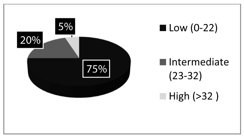
There were 75.00%, (n=30) had SYNTAX score 0 to 22 (low), 20.00%,(n=08) had 23 to 32 (intermediate) and 5.00%,(n=02) had >32 (high) according to extend of involvement of coronary arteries in the angiogram amongst the diseased group Figure 1.
CIMT in different SYNTAX score group
The mean CIMT for right and left carotid arteries were 0.62±0.11mm and 0.65±0.13mm respectively Higher CIMT value was noted among the patients with SYNTAX score ≥ 23 (7.55±3.01) than patients with SYNTAX score ≤ 22 (6.87±1.93) in the diseased group, the difference was not statistically significant (p=0.087) Table 2.
Different ABPM patterns in different SYNTAX score groups
In patients with low SYNTAX score had 45%, (n=18) abnormal dipping pattern in ABPM whereas only 12.5%, (n=05) in the intermediate to high score group showed this pattern, which was statistically significant (X2=30.7, p=0.004).
When considering the diseased group, the group with low score (SYNTAX ≤ 22) vs intermediate to high score (SYNTAX ≥ 23) had their invasive BP derived Pulse Pressure [PP] (57.70±27.60 vs 62.40±15.01mmHg, p=0.201), Fractional pulse pressure [PP/Mean BP] (0.60±0.21 vs 0.67±0.19, p=0.850) and Pulsatility index [PP/Diastolic BP](0.81±0.38 vs.0.88±0.32, p=0.707) respectively denoting that PP, fractional PP or PI did not differ among different syntax score groups.
Invasive blood pressure indices amongst diseased vs. control group
In the diseased sample, the invasive BP derived Systolic BP (SBP), Diastolic BP (DBP), Mean BP, Pulse Pressure (PP) and Pulsatillity Index (PI) was 117.50±15.80mmHg, 67.03±7.78mmHg, 83.85±9.71mmHg, 50.48±11.49 and 0.76±0.17 respectively.
In the control sample, the invasive BP derived Systolic BP (SBP), Diastolic BP (DBP), Mean BP, Pulse Pressure (PP) and Pulsatility Index (PI) was 111.05±9.47mmHg, 62.80±7.14mmHg, 78.88±7.30mmHg, 48.25±06.91 and 0.78±0.15 respectively (Table 3).
Thus statistically, the invasively derived pulsatility index showed no significant difference between the diseased and control group (0.76±0.17 and 0.78±0.15, p=0.318).
Mean CIMT diseased vs. control
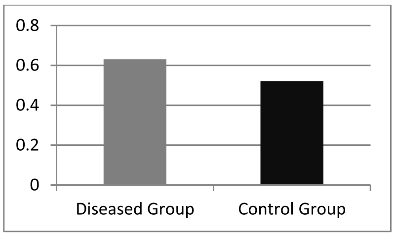
The mean CIMT in the diseased group and control group were 0.63±0.10 mm and 0.52±0.19 mm respectively (p=0.196) Figure 2.
Although the mean CIMT showed a lower value in the control group with no CAD, the difference was not statistically significant.
Diastolic dysfunction (E/e’) patients vs. controls
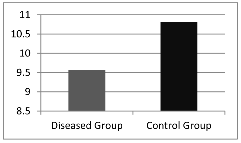
The E/e’ in the diseased group and control group were 9.56±5.40 and 10.81±3.22 respectively (p=0.372) which was not statistically significant Figure 3.
Ambulatory BP comparison
In addition, the ambulatory BP derived atherogenic index did not have any correlation with invasively derived PI both in the diseased group (r=0.005, p=0.658) and control group (r=0.003, p=0.554).
Discussion
Our study has shown the association between the pulsatile components of afterload also known as the aortic pulsatility, which is derived by the pulsatility index on invasive BP measurements and the left ventricular diastolic dysfunction. Studies in animal models have shown that increased aortic afterload results in LV diastolic dysfunction, characterized by impaired LV relaxation and increased LV end-diastolic pressures [13]. Consequently, elevated central aortic blood pressure may contribute to LV diastolic dysfunction in humans as well. This becomes clinically significant given the high prevalence of LV diastolic dysfunction in patients with hypertension, type 2 diabetes mellitus, obesity, and other related diseases [13,14]. However, studies investigating the association between central aortic pressures and diastolic function have been limited. The present study was designed to test that the pulsatile component of central aortic pressure significantly contributes to left ventricular diastolic function. The clinical importance of this finding rests in the potential for improving diastolic function (both LV relaxation and LV filling pressures) by lowering central blood pressure, an untested hypothesis that requires further study. The results of the present study may provide mechanistic insight regarding the association between pulse pressure and end-organ damage, particularly diastolic heart failure
It has been previously shown that increased afterload and central artery reflected waves contribute to LV hypertrophy. LV hypertrophy is itself strongly associated with LV diastolic filling pressures [14]. The results of the present study extend upon these findings by demonstrating that the pulsatile components of afterload contribute to LV filling pressure independent of LV mass.
Sharman et. al., investigated the relationship between arterial wave properties and diastolic function and found an association between central aortic pulse pressure and diastolic function [14]. However, this analysis was limited to a diabetic cohort and found that the partial contribution of PI to E/e’ in multivariate analysis was rather small. The present study not only validated the finding that PI contributes to high E/e’ in diabetics and non-diabetics, but also in subjects with other comorbidities such as hypertension, dyslipidaemia, and smoking. Analysis of Myocardial Performance Index (MPI) would have been a better predictor of global LV function since it is considered as a sum of an index reflecting LV systolic function as well as an index reflecting LV diastolic function [15]. The MPI is derived by dividing the sum of isovolumetric relaxation time and isovolumetric contraction time by ejection time. Ejection time is usually obtained by measuring the timing of the transaortic valve flow velocity or LV outflow velocity by continuous-wave Doppler. This measurement is observer dependent and has substantial intra-observer as well as inter-observer variability on repeated estimation [15]. Thus MPI was not in cooperated as a measurement of interest in our study. However, in future with the advent of real time three dimensional (3D) echocardiography, precise measurement of ejection time is possible and will then be a valuable tool to assess global LV function and risk stratify cardiovascular morbidity.
One of the aims of our study was to see whether increased Carotid Intima-Media Thickness (CIMT) has an association with the extent coronary artery disease and its severity. The severity and extent of CAD is measured by the SYNTAX scoring system which has world-wide acceptance [12]. Traditionally CIMT has been associated with coronary artery disease and stroke; However, emerging strong evidence from recent meta-analyses and systematic reviews suggest that its clinical usefulness may be limited because the variable CIMT measurements inconsistent with underlying CAD [16]. This is also reflected in our study where CIMT did not show significant difference between patients with CAD and control group. The measurements between the three SYNTAX score groups amongst patients with angiographically evident CAD was not significant either. These finding can partly be explained by the fact that CIMT has association with other traditional risk factors namely age, hypertension, dyslipidaemia, abdominal obesity, smoking and glucose intolerance. The impact of these risk factors on CIMT has previously been well described [17].
Interestingly, A significant proportion of patients with abnormal dipping pattern in Ambulatory Blood Pressure Monitoring (ABPM) was observed in the low SYNTAX score group compared to intermediate and high score group. Abnormal dipping pattern or reverse dipping in ABPM is an independent cardiovascular CV risk factor. However, its impact on CV mortality has not been investigated sufficiently in clinical practice for it to be considered a standard parameter [18-20].
Conclusion
In patients with CAD the diastolic dysfunction which is reflected by E/e’ positively correlated with invasively derived PI which indicate incipient arterial stiffness. Therefore E/e’ could be used as a non-invasive reproducible tool to assess arterial stiffness indirectly. CIMT showed a higher value in patients with high SYNTAX score group compared to low score group and control group without CAD, reflecting the generalized atherosclerotic process in the studied arteries although the values were not significant statistically. Statistically significant abnormal dipping pattern was observed in low SYNTAX group compared to high group which demonstrates the difference in BP variability and haemodynamic diversity amongst CAD patients with different atherosclerotic disease burden, categorized by means of SYNTAX score. These findings need further detailed evaluation to estimate the extent and severity of CAD in a non-invasive manner.
Limitations
The present study was observational and cross-sectional, and thus causality cannot be accurately determined; furthermore, long-term clinical events and outcomes were not collected. Left Ventricular (LV) diastolic function (LV filling pressure and LV relaxation) was assessed by echocardiographic tissue Doppler study rather than by invasive measurement which is the gold-standard method. LV systolic function estimation by M-mode echo may be erroneous and the 3D assessment of left ventricular ejection fraction would have been a more precise estimation of the true LV systolic function. Some patients may have sleep apnoea, which has been associated with increased aortic stiffness and diastolic dysfunction [14].
We thank our patients at cardiology unit Kandy, Sri Lanka and the staff of the cardiology unit, for their contribution towards the success of this study.
- World Health Organization (2016) Cardiovascular diseases (CVDs) Fact sheet. Link: http://bit.ly/3naY0nk
- Zafari AM, Abdou MH (2016) ‘Myocardial Infarction’. emedicine.medscape.com. Link: http://bit.ly/2XkHkPP
- Schönenberger E, Schnapauff D, Teige F, Laule M, Hamm B, et al. (2007) Patient Acceptance of Noninvasive and Invasive Coronary Angiography. Plos One 2: e246. Link: http://bit.ly/3rTUVf0
- García-Ortiz L, Recio-Rodríguez JI, Martín-Cantera C, Martín-Cantera C, Gómez-Arranz A, et al. (2010) Physical exercise, fitness and dietary pattern and their relationship with circadian blood pressure pattern, augmentation index and endothelial dysfunction biological markers: EVIDENT study protocol. BMC Public Health 10: 233. Link: http://bit.ly/3b3Ig2O
- McEniery CM, Cockcroft JR, Roman MJ, Franklin SS, Wilkinson IB (2014) Central blood pressure: current evidence and clinical importance. Eur Heart J 35: 1719-1725. Link: http://bit.ly/3nk1nbH
- Oakagomi A, Okada S, Shoji T, Kobayashi Y (2005) Aortic pulsatility assessed by an oscillometric method is associated with coronary atherosclerosis in elderly people. Blood Pressure 14: 293-297. Link: http://bit.ly/355wD7Z
- Guray Y, Guray U, Altay H, Cay S, Yilmaz MB, et al. (2005) Aortic pulse pressure and aortic pulsatility are associated with angiographic coronary artery disease in women. Blood Press 14: 293-297. Link: http://bit.ly/3rRfI2M
- Kim HL, Seo JB, Chung WY, Kim SH, Kim MA, et al. (2015) Association between invasively measured central aortic pressure and left ventricular diastolic function in patients undergoing coronary angiography. Am J Hypertens 28: 393-400. Link: http://bit.ly/3n9okOn
- Kim EJ, Park CG, Park JS, Suh SY, Choi CU, et al. (2007) Relationship between blood pressure parameters and pulse wave velocity in normotensive and hypertensive subjects: invasive study. J Hum Hypertens 21: 141-148. Link: http://bit.ly/3nckwMD
- Hillis GS, Moller JE, Pellikka PA, Gersh BJ, Wright RS, et al. (2004) Noninvasive estimation of left ventricular filling pressure by E/e’ is a powerful predictor of survival after acute myocardial infarction. J Am Coll Cardiol 43: 360-367. Link: http://bit.ly/2JH0aND
- Bella JN, Palmieri V, Roman MJ, Liu JE, Welty TK, et al. (2002) Mitral ratio of peak early to late diastolic filling velocity as a predictor of mortality in middle-aged and elderly adults: the Strong Heart Study. Circulation 105: 1928-1933. Link: http://bit.ly/38g24yt
- Sianos G, Morel MA, Kappetein AP, Morice MC, Colombo A, et al. (2005) The syntax score: an angiographic tool grading the complexity of coronary artery disease. EuroIntervention 1: 219-227. Link: http://bit.ly/3b5uwF1
- Kim HL, Im MS, Seo JB, Chung WY, Kim SH, et al. (2013) The association between arterial stiffness and left ventricular filling pressure in an apparently healthy Korean population. Cardiovasc Ultrasound 11: 2. Link: http://bit.ly/392juhb
- Mottram PM, Haluska BA, Leano R, Carlier S, Case C, et al. (2005) Relation of arterial stiffness to diastolic dysfunction in hypertensive heart disease. Heart 91: 1551-1556. Link: http://bit.ly/3odS0eD
- Carluccio E, Biagioli P, Alunni G, Murrone A, Zuchi C, et al. (2012) Improvement of myocardial performance (Tei) index closely reflects intrinsic improvement of cardiac function: assessment in revascularized hibernating myocardium. Echocardiography 29: 298-306. Link: http://bit.ly/3pMR1CB
- Granér M, Varpula M, Kahri J, Salonen RM, Nyyssönen K, et al. (2006) Association of carotid intima-media thickness with angiographic severity and extent of coronary artery disease. Am J Cardiol 97: 624-629. Link: http://bit.ly/3n9DK5d
- Gariepy J, Denarie N, Chironi G, Salomon J, Levenson J, et al. (2000) Gender difference and the influence of smoking on arterial wall thickness. Atherosclerosis 153: 139-145. Link: http://bit.ly/3b1H3ct
- Routledge FS, McFetridge-Durdle JA, Dean CR (2007) Night-time blood pressure patterns and target organ damage: a review. Can J Cardiol 23: 132-138. Link: http://bit.ly/3rSnhWU
- Roman MJ, Devereux RB, Kizer JR, Lee ET, Galloway JM, et al. (2007) Central Pressure More Strongly Relates to Vascular Disease and Outcome Than Does Brachial Pressure (The Strong Heart Study). Hypertension 50: 197-203. Link: http://bit.ly/3hFxLE7
- Al Saffar HB, Al Ghizzi HJ (2009) The Ankle- Brachial Pressure Index as A Predictor of Coronary Artery Disease Severity. Journal of the Faculty of Medicine Baghdad 51: 254-258. Link: http://bit.ly/2X4kiMV


